From Surf Wiki (app.surf) — the open knowledge base
VDAC1
Protein-coding gene in the species Homo sapiens
Protein-coding gene in the species Homo sapiens
Voltage-dependent anion-selective channel 1 (VDAC-1) is a beta barrel protein that in humans is encoded by the VDAC1 gene located on chromosome 5. It forms an ion channel in the outer mitochondrial membrane (OMM) and also the outer cell membrane. In the OMM, it allows ATP to diffuse out of the mitochondria into the cytoplasm. In the cell membrane, it is involved in volume regulation. Within all eukaryotic cells, mitochondria are responsible for synthesis of ATP among other metabolite needed for cell survival. VDAC1 therefore allows for communication between the mitochondrion and the cell mediating the balance between cell metabolism and cell death. Besides metabolic permeation, VDAC1 also acts as a scaffold for proteins such as hexokinase that can in turn regulate metabolism.
This protein is a voltage-dependent anion channel and shares high structural homology with the other VDAC isoforms (VDAC2 and VDAC3), which are involved in the regulation of cell metabolism, mitochondrial apoptosis, and spermatogenesis. Over expression and misregulation of this pore could lead to apoptosis in the cell leading to a variety of diseases within the body. In particular, since VDAC1 is the major calcium ion transport channel, its dysfunction is implicated in cancer, Parkinson's (PD), and Alzheimer's disease. In addition, recent studies have shown that an over expression within the VDAC1 protein is linked to Type 2 Diabetes. Lund University released a study that demonstrated the effects of blocking VDAC1 over expression can prevent the spread of Type 2 Diabetes.[[File:MVDAC1 structure.png|thumb|mVDAC1 top down view with alpha helix in the center of the pore.]]
Structure
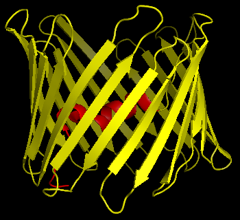
The three VDAC isoforms (VDAC1, VDAC2, and VDAC3) have highly conserved DNA sequences as well as 3D structures forming a wide β-barrel structure, inside of which the alpha helical N-terminal segment resides to partially close the pore. VDAC1's structure was solved by 3 independent labs by x-ray crystallography, Nuclear Magnetic Resonance (NMR) spectroscopy, or a combination of both. Two of these structural studies were used to determine human VDAC1 (hVDAC1) structure while X-ray crystallography was used to solve murine VDAC1 (mVDAC1) structure that differs from hVDAC1 by only two residues. These determined structures aligned with earlier circular dichroism studies that predicted the presence of alpha helix and β-strand domains.
Structural analysis of mVDAC1's structure showed a barrel-like channel composed of 19 amphipathic β-strands, with the N-terminus and C-terminus both facing towards the inter membrane space of the mitochondrion. β-strands are connected via loops and are arranged in an anti-parallel pattern with the exception of β-strands 1 and 19 which are parallel. The pore has a height of 40 Ẳ, spans a distance of 27 Ẳ by 20 Ẳ at the openings and tapers down to 20 Ẳ by 14 Ẳ at the N-terminal α-helix segment in the open state. The closed state conformation has yet to be isolated and determined. Additionally, the N-terminus has an alpha helical segment that is held to the inside wall of the pore by hydrophobic interactions with residues on β-sheets 8-18. This N-terminus can serve as a scaffold for the movement of ions or attachment of proteins. One such example is seen as it is the docking site for HK1 binding. A significant residue to point out is the glutamate located at the 73rd residue on the amino acid chain (E73). This residue is found in VDAC1 and VDAC2 but not VDAC3. The side chain of this charged residue points into the phospholipid bilayer which would normally cause repulsive forces to occur. E73 however, has been implicated in VDAC1 function and interaction.
Function
VDAC1 belongs to the mitochondrial porin family and is expected to share similar biological functions to the other VDAC isoforms. Of the three isoforms, VDAC1 is the main calcium ion transport channel in mitochondria and the most abundantly transcribed. VDAC1 is involved in cell metabolism by transporting ATP and other small metabolites across the outer mitochondrial membrane (OMM) allowing regulation of the TCA cycle and, by extension, reactive oxygen species (ROS) production. In yeast cells, ROS accumulate under conditions of oxidative stress, which results in impaired mitochondrial function and a "petite" phenotype. However, petite yeast cells exhibit a longer lifespan than wild-type cells and indicate a protective function by VDAC1 in similar circumstances, such as aging.
Voltage gating
VDAC1 allows for the conductance of molecules into and out of the mitochondrion. Its permeability is dependent on VDAC1's conformational state which is determined by voltage. At low voltage (10mV), the pore is in an "open" state where the channel is weakly anion selective and allows for a greater flux of metabolites. Because of the large pore size, metabolic gating under saturated ATP conditions reveal a transport of 2,000,000 ATP/second and a transport of 10,000 ATP under physiological conditions. At a higher voltage in the positive or negative direction (30mV), the pore is in a "closed" state and is weakly cation selective allowing for less metabolites to be transported. The flux of metabolites can be seen as negligible. This change in states is mediated by a conformational change in the protein that has yet to be discovered. Since the alpha helical N-terminus segment is located in the center of the pore, it is ideally situated for metabolic gating. This lead researchers to believe that the Alpha helix was a key contributor to determining the conformational states. However, more recent studies have shown the N-terminal is unnecessary for proper voltage gating and therefore suggest the flexible beta barrel as the mechanism of conformational change.
Oligomerization
Atomic Force Microscopy (AFM) revealed the presence of VDAC1 monomers as well as dimers and larger oligomers showcasing the interaction of the pore with itself, however, dimers are more frequent. hVDAC1 in particular has been shown to arrange in parallel dimers leading to increased permeability of the pore. The glutamate located at the 73rd position on VDAC1 has also been shown to play a role in oligomerization when in the presence of calcium. VDACs can also oligomerize to form part of the mitochondrial permeability transition pore (MPTP) and, thus, facilitate cytochrome C release, leading to apoptosis. VDACs have also been observed to interact with pro- or antiapoptotic proteins, such as Bcl-2 family proteins and kinases, and so may contribute to apoptosis independently from the MPTP.
Clinical significance
The voltage dependent anion channels all function in ion and metabolite transport although their physiological roles are different. Because of their role, dysfunction of the channels can lead to various diseases. VDAC1 has been implicated in cancer through its interactions with the antiapoptotic family of proteins, Bcl-2 proteins, particularly Bcl-xl, and Mcl-1, which are overexpressed during cancer. These two Bcl-2 proteins interact with VDAC1 to regulate calcium ion transport across the OMM and, ultimately, ROS production. While high levels of ROS induce cell death, non-lethal levels interfere with signal transduction pathways that can then promote cell proliferation, migration, and invasion in cancer cells. Moreover, VDAC1 overexpression has been associated with increased apoptotic response and anti-cancer drugs and treatment efficacy, further supporting VDAC1 as a therapeutic target for cancer treatment.
VDAC1's function in calcium ion transport has also been linked to neurodegenerative diseases. In PD, VDAC1 increases calcium ion levels within the mitochondria, resulting in increased mitochondrial permeability, disrupted mitochondrial membrane potential, elevated ROS production, cell death, and neuronal degeneration. VDAC1 has been shown to interact with Amyloid β (Aβ) leading to increased conductance of the channel and eventually apoptosis of the cell. In experimental models of Huntington's disease, treatment with the VDAC-targeting compound olesoxime improved disease-associated phenotypes, possibly by modulating calcium homeostasis and the activity of calcium-dependent calpain proteases.
Interactions
VDAC1 acts as a scaffold for many proteins as well as allows for the flux of ions and metabolites through interactions within the pore.
A major metabolite that moves through this channel is ATP. A low affinity binding site used for fast transport of this molecule was discovered by the Markov state modeling approach. It was shown that ATP binds to multiple basic residues within the pore sequentially, in essence moving through the channel.
VDAC1 has also been shown to interact with:
- BCL2-like 1,
- Bcl-2-associated X protein,
- DYNLT3,
- Gelsolin,
- PRKCE,
- HK1
- Parkin
- eNOS
- Mcl-1
- HK
- ATP
- GSK-3
References
References
- (March 1994). "Human genes encoding the voltage-dependent anion channel (VDAC) of the outer mitochondrial membrane: mapping and identification of two new isoforms". Genomics.
- "Entrez Gene: VDAC1 voltage-dependent anion channel 1".
- (February 2011). "Voltage-dependent anion channel 2 modulates resting Ca²+ sparks, but not action potential-induced Ca²+ signaling in cardiac myocytes". Cell Calcium.
- (November 2012). "Voltage-dependent anion channel-2 interaction with nitric oxide synthase enhances pulmonary artery endothelial cell nitric oxide production". American Journal of Respiratory Cell and Molecular Biology.
- (July 2003). "VDAC2 inhibits BAK activation and mitochondrial apoptosis". Science.
- (February 2012). "Critical role for voltage-dependent anion channel 2 in infectious bursal disease virus-induced apoptosis in host cells via interaction with VP5". Journal of Virology.
- (October 2014). "Mcl-1 promotes lung cancer cell migration by directly interacting with VDAC to increase mitochondrial Ca2+ uptake and reactive oxygen species generation". Cell Death & Disease.
- (September 2014). "Abnormal alpha-synuclein reduces nigral voltage-dependent anion channel 1 in sporadic and experimental Parkinson's disease". Neurobiology of Disease.
- (December 2015). "The Voltage-dependent Anion Channel 1 Mediates Amyloid β Toxicity and Represents a Potential Target for Alzheimer Disease Therapy". The Journal of Biological Chemistry.
- (January 2019). "Preserving Insulin Secretion in Diabetes by Inhibiting VDAC1 Overexpression and Surface Translocation in β Cells". Cell Metabolism.
- (2014). "Charged residues distribution modulates selectivity of the open state of human isoforms of the voltage dependent anion-selective channel". PLOS ONE.
- (October 2008). "Structure of the human voltage-dependent anion channel". Proceedings of the National Academy of Sciences of the United States of America.
- (August 2008). "Solution structure of the integral human membrane protein VDAC-1 in detergent micelles". Science.
- (November 2008). "The crystal structure of mouse VDAC1 at 2.3 A resolution reveals mechanistic insights into metabolite gating". Proceedings of the National Academy of Sciences of the United States of America.
- (April 2007). "Correct folding of the beta-barrel of the human membrane protein VDAC requires a lipid bilayer". Journal of Molecular Biology.
- (June 2012). "The role of VDAC in cell death: friend or foe?". Biochimica et Biophysica Acta (BBA) - Biomembranes.
- (December 2013). "The voltage-dependent anion selective channel 1 (VDAC1) topography in the mitochondrial outer membrane as detected in intact cell". PLOS ONE.
- (March 2012). "Affixing N-terminal α-helix to the wall of the voltage-dependent anion channel does not prevent its voltage gating". The Journal of Biological Chemistry.
- (July 2010). "Swapping of the N-terminus of VDAC1 with VDAC3 restores full activity of the channel and confers anti-aging features to the cell". FEBS Letters.
- (July 2013). "The role of calcium in VDAC1 oligomerization and mitochondria-mediated apoptosis". Biochimica et Biophysica Acta (BBA) - Molecular Cell Research.
- (May 2004). "Identification of the hypoxia-inducible factor 1 alpha-responsive HGTD-P gene as a mediator in the mitochondrial apoptotic pathway". Molecular and Cellular Biology.
- (May 2025). ["Characterization of human VDAC isoforms: a peculiar function for VDAC3?"](https://www.openaccessrepository.it/record/135813 }}{{dead link). Biochimica et Biophysica Acta (BBA) - Bioenergetics.
- (May 1997). "VDAC channels mediate and gate the flow of ATP: implications for the regulation of mitochondrial function". Biophysical Journal.
- (July 2007). "The supramolecular assemblies of voltage-dependent anion channels in the native membrane". Journal of Molecular Biology.
- (October 2014). "Ca(2+)-mediated regulation of VDAC1 expression levels is associated with cell death induction". Biochimica et Biophysica Acta (BBA) - Molecular Cell Research.
- (2016-01-01). "The calpain-suppressing effects of olesoxime in Huntington's disease". Rare Diseases.
- (October 2019). "Olesoxime in neurodegenerative diseases: Scrutinising a promising drug candidate". Biochemical Pharmacology.
- (December 2015). "Olesoxime suppresses calpain activation and mutant huntingtin fragmentation in the BACHD rat". Brain.
- (July 2014). "Structure-guided simulations illuminate the mechanism of ATP transport through VDAC1". Nature Structural & Molecular Biology.
- (March 2005). "Specific cleavage of Mcl-1 by caspase-3 in tumor necrosis factor-related apoptosis-inducing ligand (TRAIL)-induced apoptosis in Jurkat leukemia T cells". The Journal of Biological Chemistry.
- (June 2003). "Identification of the protein-protein contact site and interaction mode of human VDAC1 with Bcl-2 family proteins". Biochemical and Biophysical Research Communications.
- (March 2000). "BH4 domain of antiapoptotic Bcl-2 family members closes voltage-dependent anion channel and inhibits apoptotic mitochondrial changes and cell death". Proceedings of the National Academy of Sciences of the United States of America.
- (June 1999). "Bcl-2 family proteins regulate the release of apoptogenic cytochrome c by the mitochondrial channel VDAC". Nature.
- (September 2002). "Voltage-dependent anion-selective channel (VDAC) interacts with the dynein light chain Tctex1 and the heat-shock protein PBP74". The International Journal of Biochemistry & Cell Biology.
- (October 2000). "Human gelsolin prevents apoptosis by inhibiting apoptotic mitochondrial changes via closing VDAC". Oncogene.
- (May 2003). "Protein kinase Cepsilon interacts with and inhibits the permeability transition pore in cardiac mitochondria". Circulation Research.
- (November 2012). "Voltage-dependent anion channels (VDACs) recruit Parkin to defective mitochondria to promote mitochondrial autophagy". The Journal of Biological Chemistry.
- (December 2013). "ATP transport through VDAC and the VDAC-tubulin complex probed by equilibrium and nonequilibrium MD simulations". Biochemistry.
- (January 2013). "Glycogen synthase kinase 3-mediated voltage-dependent anion channel phosphorylation controls outer mitochondrial membrane permeability during lipid accumulation". Hepatology.
This article was imported from Wikipedia and is available under the Creative Commons Attribution-ShareAlike 4.0 License. Content has been adapted to SurfDoc format. Original contributors can be found on the article history page.
Ask Mako anything about VDAC1 — get instant answers, deeper analysis, and related topics.
Research with MakoFree with your Surf account
Create a free account to save articles, ask Mako questions, and organize your research.
Sign up freeThis content may have been generated or modified by AI. CloudSurf Software LLC is not responsible for the accuracy, completeness, or reliability of AI-generated content. Always verify important information from primary sources.
Report