From Surf Wiki (app.surf) — the open knowledge base
Urethra
Tube that connects the urinary bladder to the external urethral orifice
Tube that connects the urinary bladder to the external urethral orifice
| Field | Value |
|---|---|
| Name | Urethra |
| Latin | urethra feminina (female); urethra masculina (male) |
| Greek | οὐρήθρα |
| Image | Female and Male Urethra.jpg |
| Caption | The urethra transports urine from the bladder to the outside of the body. This image shows (a) a human female urethra and (b) a human male urethra. |
| Width | 220 |
| Precursor | Urogenital sinus |
| Artery | Inferior vesical artery |
| Middle rectal artery | |
| Internal pudendal artery | |
| Vein | Inferior vesical vein |
| Middle rectal vein | |
| Internal pudendal vein | |
| Nerve | Pudendal nerve |
| Pelvic splanchnic nerves | |
| Inferior hypogastric plexus | |
| Lymph | Internal iliac lymph nodes |
| Deep inguinal lymph nodes |
Middle rectal artery Internal pudendal artery Middle rectal vein Internal pudendal vein Pelvic splanchnic nerves Inferior hypogastric plexus Deep inguinal lymph nodes
The urethra (: urethras or urethrae) is the tube that transports urine from the urinary bladder to the external urethral meatus of the penis or vulva in placental mammals. In males, the urethra transports semen through the penis during ejaculation.
The external urethral sphincter is a striated muscle that allows voluntary control over urination. The internal sphincter, formed by the involuntary smooth muscles lining the bladder neck and urethra, is innervated by the sympathetic division of the autonomic nervous system and is found both in males and females.
Structure
The urethra is a fibrous and muscular tube which connects the urinary bladder to the external urethral meatus. Its length differs between the sexes, because it passes through the penis in males.
Male
In the human male, the urethra is on average 18 to long and opens at the end of the external urethral meatus.
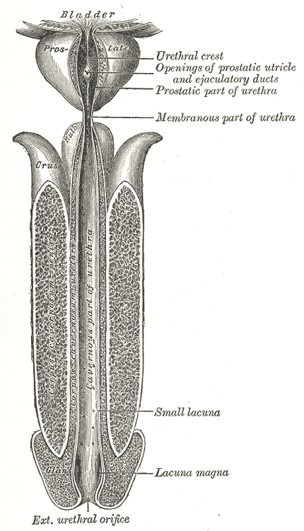
The urethra is divided into four parts in men, named after the location:
| Region | Description | Epithelium |
|---|---|---|
| Pre-prostatic urethra | This is the intramural part of the urethra surrounded by the internal urethral sphincter and varies between 0.5 and 1.5 cm in length depending on the fullness of the bladder. | Transitional |
| Prostatic urethra | Crosses through the prostate gland. There are several openings at the posterior wall: | Transitional |
| Membranous urethra | A short (1 or 2 cm) portion passing through the external urethral sphincter. This is the narrowest part of the urethra. It is located in the deep perineal pouch. The bulbourethral glands (Cowper's gland) are found posterior to this region but open in the spongy urethra. | Pseudostratified columnar |
| Spongy urethra (or penile urethra) | Runs along the length of the penis on its ventral (underneath) surface. It is about 15 to 25 cm in length, with steady diameter of 6 mm, and travels through the corpus spongiosum. The ducts from the urethral gland (gland of Littré) enter here. The openings of the bulbourethral glands are also found here. Some textbooks will subdivide the spongy urethra into two parts, the bulbous and pendulous urethra. The urethral lumen runs effectively parallel to the penis, except at the narrowest point, the external urethral meatus, where it is vertical. This produces a spiral stream of urine and has the effect of cleaning the external urethral meatus. The lack of an equivalent mechanism in the female urethra partly explains why urinary tract infections occur so much more frequently in females. | Pseudostratified columnar – proximallyStratified squamous – distally |
There is inadequate data for the typical length of the male urethra; however, a study of 109 men showed an average length of 22.3 cm (SD = 2.4 cm), ranging from 15 cm to 29 cm.
The urethra in male placental mammals is typically longer than in females.
Female
In the human female, the urethra is about 4 cm long, having 6 mm diameter, and exits the body between the clitoris and the vaginal opening, extending from the internal to the external urethral orifice. The meatus is located below the clitoris. It is placed behind the symphysis pubis, embedded in the anterior wall of the vagina, and its direction is obliquely downward and forward; it is slightly curved with the concavity directed forward. The proximal two-thirds of the urethra is lined by transitional epithelial cells, while the distal third is lined by stratified squamous epithelial cells.
Between the superior and inferior fascia of the urogenital diaphragm, the female urethra is surrounded by the urethral sphincter.
The urethra in female placental mammals is typically shorter than in the male.
Microanatomy
The cells lining the urethra (the epithelium) going all over transitional cells as it exits the bladder, which are variable layers of flat to cuboidal cells that change shape depending on whether they are compressed by the contents of the urethra. Further along the urethra there are pseudostratified columnar and stratified columnar epithelia. The lining becomes multiple layers of flat cells near the end of the urethra, which is the same as the external skin around it.
There are small mucus-secreting urethral glands, as well as bulbo-urethral glands of Cowper, that secrete mucous acting to lubricate the urethra.
The urethra consists of three coats: muscular, erectile, and mucous, the muscular layer being a continuation of that of the bladder.
Blood and nerve supply and lymphatics
Somatic (conscious) innervation of the external urethral sphincter is supplied by the pudendal nerve.
Development
In the developing embryo, at the hind end lies a cloaca. This, over the fourth to the seventh week, divides into a urogenital sinus and the beginnings of the anal canal, with a wall forming between these two inpouchings called the urorectal septum. The urogenital sinus divides into three parts, with the middle part forming the urethra; the upper part is largest and becomes the urinary bladder, and the lower part then changes depending on the biological sex of the embryo. The cells lining the urethra (the epithelium) come from endoderm, whereas the connective tissue and smooth muscle parts are derived from mesoderm.
After the third month, urethra also contributes to the development of associated structures depending on the biological sex of the embryo. In the male, the epithelium multiples to form the prostate. In the female, the upper part of the urethra forms the urethra and paraurethral glands.
Function
Urination
The urethra is the vessel through which urine exits the bladder. During urination, the urethra's smooth muscle relaxes as the bladder contracts to expel urine in a pressurized stream. Following this, the urethra re-establishes muscle tone by contracting the smooth muscle layer, and the bladder returns to a relaxed, quiescent state. Urethral smooth muscle cells are mechanically coupled to each other to coordinate mechanical force and electrical signaling in an organized, unitary fashion.
Ejaculation
The male urethra is the conduit for semen during orgasm. Urine is removed before ejaculation by pre-ejaculate fluid – called Cowper's fluid – from the bulbourethral gland.
Clinical significance

Infection of the urethra is urethritis, which often causes purulent urethral discharge.
Cancer can also develop in the lining of the urethra. When cancer is present, the most common symptom in an affected person is blood in the urine; a physical medical examination may be otherwise normal, except in late disease. Cancer of the urethra is most often due to cancer of the cells lining the urethra, called transitional cell carcinoma, although it can more rarely occur as a squamous cell carcinoma if the type of cells lining the urethra have changed, such as due to a chronic schistosomiasis infection. Investigations performed usually include collecting a sample of urine for an inspection for malignant cells under a microscope, called cytology, as well as examination with a flexible camera through the urethra, called urethroscopy. If a malignancy is found, a biopsy will be taken, and a CT scan will be performed of other body parts (a CT scan of the chest, abdomen and pelvis) to look for additional lesions. After the cancer is staged, treatment may involve chemotherapy.
Injury
Passage of kidney stones through the urethra can be painful. Damage to the urethra by kidney stones, chronic infection, cancer, or catheterisation can cause narrowing, called a urethral stricture. A retrograde urethrogram in which dye is injected into the urethra can reveal the location and structure of the narrowing. Other forms of imaging such as ultrasound, computed tomography and magnetic resonance imaging may also provide further details.
Injuries to the urethra (e.g., from a pelvic fracture)
Foreign bodies in the urethra are uncommon, but there have been medical case reports of self-inflicted injuries, a result of insertion of foreign bodies into the urethra such as an electrical wire.
Other
Hypospadias and epispadias are abnormal developments of the male urethra where the meatus is not at the distal end of the penis (it occurs lower than normal with hypospadias, and higher with epispadias). In a severe chordee, the urethra can develop between the penis and the scrotum.
Catheterisation
A tube called a catheter can be inserted through the urethra to drain urine from the bladder, called an indwelling urinary catheter; or, to bypass the urethra, a catheter may be directly inserted through the abdominal wall into the bladder, called a suprapubic catheter.
Other animals
In all mammals, with the exception of monotremes, and in both sexes, the urethra serves primarily to drain and excrete urine, which in mammals, collects in the urinary bladder and is released from there into the urethra. In addition, the closing mechanisms of the urethra, together with immunoglobulins, largely prevent germs from penetrating the inside of the body. In marsupials, the female's urethra empties into the urogenital sinus.
History
The word "urethra" comes from the Ancient Greek οὐρήθρα – ourḗthrā. The stem "uro" relating to urination, with the structure described as early as the time of Hippocrates. Confusingly however, at the time it was called "ureter". Thereafter, terms "ureter" and "urethra" were variably used to refer to each other thereafter for more than a millennium. It was only in the 1550s that anatomists such as Bartolomeo Eustachi and Jacques Dubois began to use the terms to specifically and consistently refer to what is in modern English called the ureter and the urethra. Following this, in the 19th and 20th centuries, multiple terms relating to the structures such as urethritis and urethrography, were coined.
Kidney stones have been identified and recorded about as long as written historical records exist. The urinary tract as well as its function to drain urine from the kidneys, has been described by Galen in the second century AD. Surgery to the urethra to remove kidney stones has been described since at least the first century AD by Aulus Cornelius Celsus.
Additional images
File:Prostatelead.jpg|Position of the urethra in males File:Gray1155 a.png|Transverse section of the penis File:Male urinary meatus.jpg|Male urethral opening on glans penis File:Skenes gland.jpg|Female urethral opening within vulval vestibule File:1116 Muscle of the Female Perineum.png|Muscles of the female perineum File:Slide12BLA.JPG|Urethra. Deep dissection. Serial cross section. File:Penis lateral cross section.jpg|Diagram which depicts the membranous urethra and the spongy urethra of a male File:Female vaginal anatomy.jpg
References
References
- Lombardi, Julian. (2012-12-06). "Comparative Vertebrate Reproduction". Springer Science & Business Media.
- Marvalee H. Wake. (15 September 1992). "Hyman's Comparative Vertebrate Anatomy". University of Chicago Press.
- Brading, Alison F.. (January 1999). "The physiology of the mammalian urinary outflow tract". Experimental Physiology.
- (2004). "Principles of Gender-specific Medicine". Gulf Professional Publishing.
- (2004). "Neurophysiology of Stress Urinary Incontinence". Reviews in Urology.
- (September 2012). "Clinical and Functional Anatomy of the Urethral Sphincter". International Neurourology Journal.
- (April 2005). "The structure and innervation of the male urethra: histological and immunohistochemical studies with three-dimensional reconstruction". Journal of Anatomy.
- (April 2007). "Functional anatomy of the female pelvic floor.". Annals of the New York Academy of Sciences.
- "Male Urethra Function & Urethra Anatomy Pictures".
- (13 October 2010). "Urethra". Chicago's Jeasuit University.
- Atlas of Human Anatomy 5th Edition, Netter.
- (2008). "The length of the male urethra". International Brazilian Journal of Urology.
- Marvalee H. Wake. (1999). "Homeopathic Care for Cats and Dogs: Small Doses for Small Animals". North Atlantic Books.
- (2016). "Gray's Anatomy: The Anatomical Basis of Clinical Practice". Elsevier Limited.
- Manual of Obstetrics. (3rd ed.). Elsevier. pp. 1-16. {{ISBN. 9788131225561.
- (2013). "Wheater's functional histology: a text and colour atlas.". Elsevier.
- (2019). "Langman's medical embryology". Wolters Kluwer.
- (Aug 2014). "Ion Channels of the Mammalian Urethra". Channels.
- (15 December 2010). "Sperm content of pre-ejaculatory fluid". Human Fertility.
- (April 2005). "A neglected gland: a review of Cowper's gland". Int. J. Androl..
- (2018). "Davidson's principles and practice of medicine". Elsevier.
- (2018). "Davidson's principles and practice of medicine". Elsevier.
- (12 December 2018). "Urethral stricture". Mayo Clinic.
- (2015-12-02). "Imaging of urethral stricture disease". Translational Andrology and Urology.
- (July 2015). "An update on urotrauma". Current Opinion in Urology.
- (2009). "Electrical wire as a foreign body in a male urethra: a case report". Journal of Medical Case Reports.
- (26 February 2020). "Urinary catheters - NHS".
- Wilson, Michaeltitle=Microbial Inhabitants of Humans: Their Ecology and Role in Health and Disease. (2005). "Microbial Inhabitants of Humans: Their Ecology and Role in Health and Disease". Cambridge University Press.
- (30 January 1987). "Reproductive Physiology of Marsupials". Cambridge University Press.
- (2010). "Uro-words making history: Ureter and urethra". The Prostate.
- (2013). "The History of Urinary Stones: In Parallel with Civilization". The Scientific World Journal.
- (2011). "The History of Urologic Surgery: From Reeds to Robotics". Urologic Nursing.
This article was imported from Wikipedia and is available under the Creative Commons Attribution-ShareAlike 4.0 License. Content has been adapted to SurfDoc format. Original contributors can be found on the article history page.
Ask Mako anything about Urethra — get instant answers, deeper analysis, and related topics.
Research with MakoFree with your Surf account
Create a free account to save articles, ask Mako questions, and organize your research.
Sign up freeThis content may have been generated or modified by AI. CloudSurf Software LLC is not responsible for the accuracy, completeness, or reliability of AI-generated content. Always verify important information from primary sources.
Report