From Surf Wiki (app.surf) — the open knowledge base
Thymoma
| Field | Value |
|---|---|
| name | Thymoma |
| image | Encapsulated thymoma.jpg |
| caption | An encapsulated thymoma (mixed lymphocytic and epithelial type) |
| field | Oncology, cardiothoracic surgery |
| onset | Adulthood |
| treatment | surgical removal, chemotherapy (in malignant cases). |
A thymoma is a tumor originating from the epithelial cells of the thymus that is considered a rare neoplasm. Thymomas are frequently associated with neuromuscular disorders such as myasthenia gravis; thymoma is found in 20% of patients with myasthenia gravis. Once diagnosed, thymomas may be removed surgically. In the rare case of a malignant tumor, radiation therapy may be used.
Signs and symptoms
A third of all people with a thymoma have symptoms caused by compression of the surrounding organs by an expansive mass. These problems may take the form of superior vena cava syndrome, dysphagia (difficulty swallowing), cough, or chest pain.
One-third of patients have their tumors discovered because they have an associated autoimmune disorder. As mentioned earlier, the most common of those conditions is myasthenia gravis (MG); 10–15% of patients with MG have a thymoma and, conversely, 30–45% of patients with thymomas have MG. Additional associated autoimmune conditions include thymoma-associated multiorgan autoimmunity, pure red cell aplasia and Good syndrome (thymoma with combined immunodeficiency and hypogammaglobulinemia). Other reported disease associations are with acute pericarditis, agranulocytosis, alopecia areata, ulcerative colitis, Cushing's disease, hemolytic anemia, limbic encephalopathy, myocarditis, nephrotic syndrome, panhypopituitarism, pernicious anemia, polymyositis, rheumatoid arthritis, sarcoidosis, scleroderma, sensorimotor radiculopathy, stiff person syndrome, systemic lupus erythematosus and thyroiditis.
One-third to one-half of all persons with thymoma have no symptoms at all, and the mass is identified on a chest X-ray or CT/CAT scan performed for an unrelated problem.
Pathology
Thymoma originates from the epithelial cell population in the thymus, and several microscopic subtypes are now recognized. There are three principal histological types of thymoma, depending on the appearance of the cells by microscopy:
- Type A if the epithelial cells have an oval or fusiform shape (less lymphocyte count);
- Type B if they have an epithelioid shape (Type B has three subtypes: B1 (lymphocyte-rich), B2 (cortical) and B3 (epithelial).);
- Type AB if the tumor contains a combination of both cell types.
Thymic cortical epithelial cells have abundant cytoplasm, vesicular nucleus with finely divided chromatin and small nucleoli and cytoplasmic filaments contact adjacent cells. Thymic medullary epithelial cells in contrast are spindle shaped with oval dense nucleus and scant cytoplasm thymoma if recapitulates cortical cell features more, is thought to be less benign.
Diagnosis
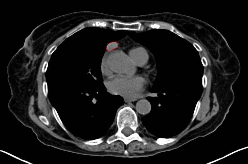
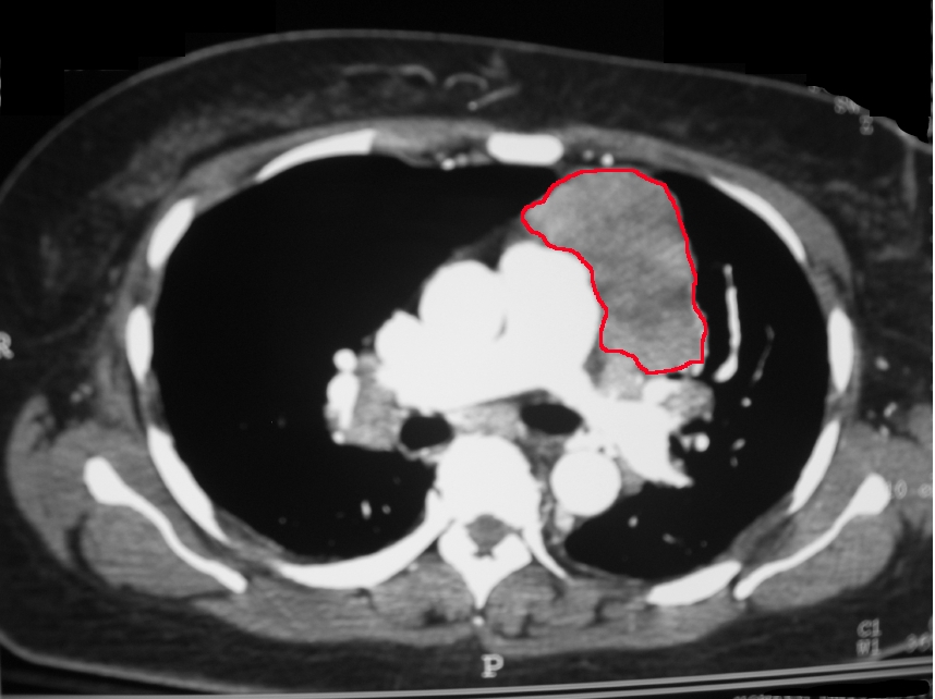
When a thymoma is suspected, a CT/CAT scan is generally performed to estimate the size and extent of the tumor, and the lesion is sampled with a CT-guided needle biopsy. Increased vascular enhancement on CT scans can be indicative of malignancy, as can be pleural deposits.
The diagnosis is made via histologic examination by a pathologist, after obtaining a tissue sample of the mass. Final tumor classification and staging is accomplished pathologically after formal surgical removal of the thymic tumor.
Selected laboratory tests can be used to look for associated problems or possible tumor spread. These include: full blood count, protein electrophoresis, antibodies to the acetylcholine receptor (indicative of myasthenia), electrolytes, liver enzymes and renal function.
Staging
The Masaoka Staging System is used widely and is based on the anatomic extent of disease at the time of surgery:
- I: Completely encapsulated
- IIA: Microscopic invasion through the capsule into surrounding fatty tissue
- IIB: Macroscopic invasion into capsule
- III: Macroscopic invasion into adjacent organs
- IVA: Pleural or pericardial implants
- IVB: Lymphogenous or hematogenous metastasis to distant (extrathoracic) sites
Treatment
Surgery is the mainstay of treatment for thymoma. If the tumor is apparently invasive and large, preoperative (neoadjuvant) chemotherapy and/or radiotherapy may be used to decrease the size and improve resectability, before surgery is attempted. When the tumor is an early stage (Masaoka I through IIB), no further therapy is necessary. Removal of the thymus in adults does not appear to induce immune deficiency. In children, however, postoperative immunity may be abnormal and vaccinations for several infectious agents are recommended. Invasive thymomas may require additional treatment with radiotherapy and chemotherapy (cyclophosphamide, doxorubicin and cisplatin). Recurrences of thymoma are described in 10-30% of cases up to 10 years after surgical resection, and in the majority of cases also pleural recurrences can be removed. Recently, surgical removal of pleural recurrences can be followed by hyperthermic intrathoracic perfusion chemotherapy or intrathoracic hyperthermic perfused chemotherapy (ITH).
Prognosis
Prognosis is much worse for stage III or IV thymomas as compared with stage I and II tumors. Invasive thymomas uncommonly can also metastasize, generally to pleura, bones, liver or brain in approximately 7% of cases.
Patients who have undergone thymectomy for thymoma should be warned of possible severe side effects after yellow fever vaccination. This is probably caused by inadequate T-cell response to live attenuated yellow fever vaccine. Deaths have been reported.
Epidemiology
The incidence of thymomas is around 0.13-0.26 per 100,000 people per year. Males are affected slightly less frequently than females. The typical age at diagnosis is in the 40s and 50s, though the age may range from six years to 83 years.
Gallery
File:Encapsulated cystic thymoma.jpg|An encapsulated cystic thymoma. File:Locally invasive circumscribed thymoma.jpg|A locally invasive circumscribed thymoma (mixed lymphocytic and epithelial, mixed polygonal and spindle). File:Thymoma B1 (2).JPG|Histopathological image of thymoma type B1. Anterior mediastinal mass surgically resected. Hematoxylin & eosin stain. File:Thymoma B1 (3) CK CAM5-2.JPG|Histopathological image of thymoma type B1. Anterior mediastinal mass surgically resected. Cytokeratin CAM5.2 immunostain. File:Thymoma type B1 (1).JPG|Histopathological image representing a noninvasive thymoma type B1, surgically resected. Hematoxylin & eosin. File:Thymoma - cytology high mag.jpg|Thymoma. FNA specimen. Field stain.
References
References
- WHO classification of tumors editorial Board. Thoracic Tumors. WHO Classification of Tumors. Lyon: IARC; 2021.
- (July 1999). "Thymoma: state of the art". Journal of Clinical Oncology.
- (May 2018). "Robbins basic pathology". Saunders/Elsevier.
- (January 2016). "Thymoma associated with autoimmune diseases: 85 cases and literature review". Autoimmunity Reviews.
- (May 2001). "Histologic typing of thymoma according to the new World Health Organization classification". Chest Surgery Clinics of North America.
- (December 1981). "Follow-up study of thymomas with special reference to their clinical stages". Cancer.
- (2016). "Pleural recurrence of thymoma: surgical resection followed by hyperthermic intrathoracic perfusion chemotherapy". Eur J Cardiothorac Surg.
- (2021). "Thoracic Tumours". World Health Organization.
This article was imported from Wikipedia and is available under the Creative Commons Attribution-ShareAlike 4.0 License. Content has been adapted to SurfDoc format. Original contributors can be found on the article history page.
Ask Mako anything about Thymoma — get instant answers, deeper analysis, and related topics.
Research with MakoFree with your Surf account
Create a free account to save articles, ask Mako questions, and organize your research.
Sign up freeThis content may have been generated or modified by AI. CloudSurf Software LLC is not responsible for the accuracy, completeness, or reliability of AI-generated content. Always verify important information from primary sources.
Report