From Surf Wiki (app.surf) — the open knowledge base
Suture (anatomy)
Fairly rigid joint between two or more hard elements of an organism

Fairly rigid joint between two or more hard elements of an organism
| Field | Value |
|---|---|
| Name | Suture |
| image | File:Human skull no text no color.svg |
| caption | Sutures of the human skull |
| Latin | sutura |
natural anatomical joints
In anatomy, a suture is a fairly rigid joint between two or more hard elements of an organism, with or without significant overlap of the elements.
Sutures are found in the skeletons or exoskeletons of a wide range of animals, in both invertebrates and vertebrates. Sutures are found in animals with hard parts from the Cambrian period to the present day. Sutures were and are formed by several different methods, and they exist between hard parts that are made from several different materials.
Vertebrate skeletons
The skeletons of vertebrate animals (fish, amphibians, reptiles, birds, and mammals) are made of bone, in which the main rigid ingredient is calcium phosphate.
Cranial sutures
The skulls of most vertebrates consist of sets of bony plates held together by cranial sutures. These sutures are held together mainly by Sharpey's fibers which grow from each bone into the adjoining one.{{cite journal
Sutures in the ankles of land vertebrates
In the type of crurotarsal ankle, which is found in crocodilians and some other archosaurs, the astragalus is fixed to the tibia by a suture, and the joint bends around a peg on the astragalus, which fits into a socket in the calcaneum.
Invertebrate exoskeletons
In molluscs
The shells of most molluscs are made of calcium carbonate (the main constituent of limestone and chalk), and of conchiolin, a protein. For more information, see Mollusc shell.
Sutures in the shells of cephalopods
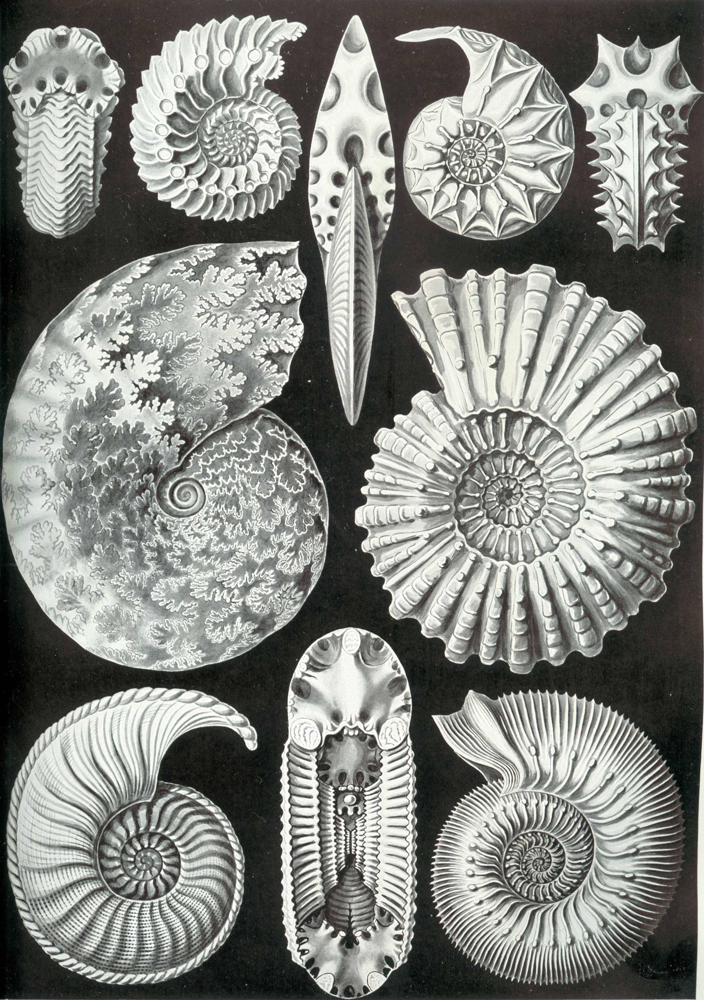
In cephalopod mollusks, which have external shells (e.g. Nautilus, ammonites), the shell is divided into compartments by septa (partitions).
The septa are joined to the external shell by sutures formed by repeated invagination (they interlock like pieces of a jigsaw puzzle). The sutures are visible from the outside and often form complex and elaborate patterns.
The suture in the shells of [[gastropod]]s{{anchor|Gastropod}}
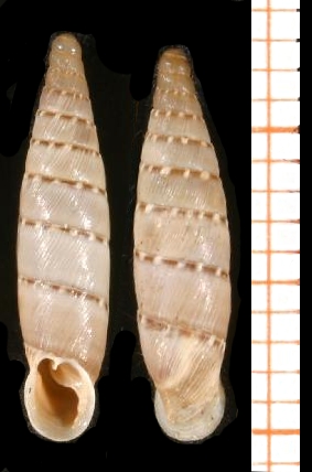
Nearly all snail shells (except for the shells of limpets, abalone, sea hares, etc.) can be visualized as a tube of increasing diameter, closed at the small end, and spirally wrapped around a central axis. For more information, see Gastropod shell.
Each complete rotation of this spirally-arranged tube is called a whorl. The whorls of a snail shell usually overlap one another, forming a spire. Where the whorls overlap, there is usually a clear (if narrow) indentation. This indentation forms a visible line, which is continuous and reaches from the apex of the shell to the aperture; this line is the suture.
Details of the suture are often useful in discriminating one species from another, for example, sometimes the suture is channeled.
The suture also provides a sort of geographic marker from which one can refer to the positioning of patterning or sculpture, where that is relevant: for example some species have a darker or lighter subsutural band on the shell.
When an angulation of the whorls occurs, the space between it and the suture above it (i.e. the abaxial edge of the sutural ramp) constitutes the area known as the "shoulder" of the shell. The shoulder angle may be simple or keeled, and may sometimes have nodes or spines
In arthropods
Sutures in the carapaces of trilobites
Main article: Facial suture (trilobite)

A trilobite's carapace consisted of calcite and calcium phosphate deposited on a lattice (framework) of chitin (a polysaccharide).
The trilobite body is divided into three major sections: a cephalon (head section) with eyes, mouthparts and sensory organs such as antennae; a thorax of multiple segments which are similar to each other; and a pygidium, or tail section.
In many species, the cephalon had sutures running from back to front round the outside edges of the eyes. These sutures divided the cephalon into three pieces.
The sutures in trilobites' cephalons were unusual because it seems their main function was to create weaknesses, which made it easy for this part of the carapace ("armor") to split when the animal needed to molt.
References
References
- (2013). "Anatomy & physiology". OpenStax.
- [http://www.palaeos.com/Vertebrates/Units/270Archosauromorpha/270.500.html Archosauromorpha: Archosauria - Palaeos] {{Webarchive. link. (2005-04-05)
- Ruppert, E.E., Fox, R.S., and Barnes, R.D.. (2004). "Invertebrate Zoology". Brooks / Cole.
- [http://www.palaeos.com/Invertebrates/Molluscs/Cyrtosoma/Cephalopoda/Cephalopoda.Glossary.html#suture Cephalopoda Glossary - Palaeos]
- "Palaeos Metazoa: Mollusca: Cyrtosoma: Cephalopoda: Glossary". Palaeos.
- Fortey, Richard A. (2000). "Trilobite!: eyewitness to evolution". Alfred Knopf.
This article was imported from Wikipedia and is available under the Creative Commons Attribution-ShareAlike 4.0 License. Content has been adapted to SurfDoc format. Original contributors can be found on the article history page.
Ask Mako anything about Suture (anatomy) — get instant answers, deeper analysis, and related topics.
Research with MakoFree with your Surf account
Create a free account to save articles, ask Mako questions, and organize your research.
Sign up freeThis content may have been generated or modified by AI. CloudSurf Software LLC is not responsible for the accuracy, completeness, or reliability of AI-generated content. Always verify important information from primary sources.
Report