From Surf Wiki (app.surf) — the open knowledge base
Striatopallidal fibres
Structure of the basal ganglia
Structure of the basal ganglia
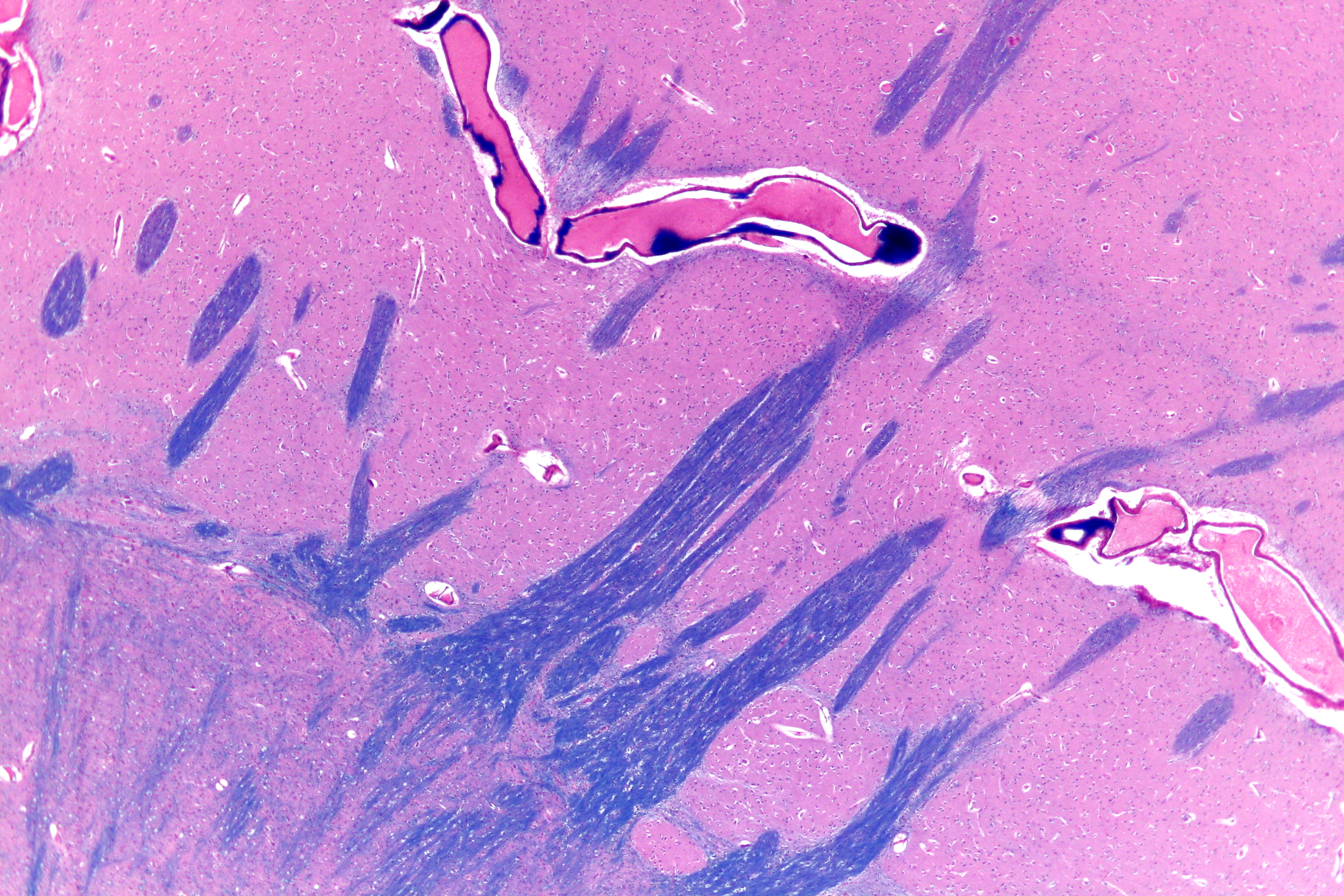
The striatopallidal fibres, also Wilson's pencils, pencil fibres of Wilson, and pencils of Wilson, are prominent myelinated fibres that connect the striatum to the globus pallidus.
Their distinctive appearance allows the putamen to be identified on light microscopy.
References
References
- (December 1996). "Neural information transferred from the putamen to the globus pallidus during learned movement in the monkey". J. Neurophysiol..
- Wilson SAK. (1914). "An experimental research into the anatomy and physiology of the corpus striatum". Brain.
- (2010). "Practical Surgical Neuropathology: A Diagnostic Approach: A Volume in the Pattern Recognition series". Churchill Livingstone.
This article was imported from Wikipedia and is available under the Creative Commons Attribution-ShareAlike 4.0 License. Content has been adapted to SurfDoc format. Original contributors can be found on the article history page.
Ask Mako anything about Striatopallidal fibres — get instant answers, deeper analysis, and related topics.
Research with MakoFree with your Surf account
Create a free account to save articles, ask Mako questions, and organize your research.
Sign up freeThis content may have been generated or modified by AI. CloudSurf Software LLC is not responsible for the accuracy, completeness, or reliability of AI-generated content. Always verify important information from primary sources.
Report