From Surf Wiki (app.surf) — the open knowledge base
Pudendal nerve
Main nerve of the perineum

Main nerve of the perineum
| Field | Value | ||
|---|---|---|---|
| Name | Pudendal nerve | ||
| Latin | nervus pudendus | ||
| Image | File:Gray542.png | ||
| Caption | Cross-section of pelvis in which nerve emerges and is visible on the right side. | ||
| Image2 | [[File:Pudendal nerve.svg | class=skin-invert-image | 250px]] |
| Caption2 | Pudendal nerve, course and branches in a male. | ||
| BranchFrom | Sacral nerves S2, S3, S4 | ||
| BranchTo | Inferior rectal nerves | ||
| perineal nerve | |||
| dorsal nerve of the penis | |||
| dorsal nerve of the clitoris |
perineal nerve dorsal nerve of the penis dorsal nerve of the clitoris
The pudendal nerve is the main nerve of the perineum. It is a mixed (motor and sensory) nerve and also conveys sympathetic autonomic fibers. It carries sensation from the external genitalia of both sexes and the skin around the anus and perineum, as well as the motor supply to various pelvic muscles, including the male or female external urethral sphincter and the external anal sphincter.
If damaged, most commonly by childbirth, loss of sensation or fecal incontinence may result. The nerve may be temporarily anesthetized, called pudendal anesthesia or pudendal block.
The pudendal canal that carries the pudendal nerve is also known by the eponymous term "Alcock's canal", after Benjamin Alcock, an Irish anatomist who documented the canal in 1836.
Structure
Origin
The pudendal nerve is paired, meaning there are two nerves, one on the left and one on the right side of the body. Each is formed as three roots immediately converge above the upper border of the sacrotuberous ligament and the coccygeus muscle.
Course and relations
The pudendal nerve passes between the piriformis muscle and coccygeus (ischiococcygeus) muscles and leaves the pelvis through the lower part of the greater sciatic foramen. It crosses over the lateral part of the sacrospinous ligament and reenters the pelvis through the lesser sciatic foramen. After reentering the pelvis, it accompanies the internal pudendal artery and internal pudendal vein upwards and forwards along the lateral wall of the ischiorectal fossa, being contained in a sheath of the obturator fascia termed the pudendal canal, along with the internal pudendal blood vessels.
Branches
Inside the pudendal canal, the nerve divides into branches, first giving off the inferior rectal nerve, then the perineal nerve, before continuing as the dorsal nerve of the penis (in males) or the dorsal nerve of the clitoris (in females).
Nucleus
The nerve is a major branch of the sacral plexus, with fibers originating in Onuf's nucleus in the sacral region of the spinal cord.
Variation
The pudendal nerve may vary in its origins. For example, the pudendal nerve may actually originate in the sciatic nerve. Consequently, damage to the sciatic nerve can affect the pudendal nerve as well. Sometimes dorsal rami of the first sacral nerve contribute fibers to the pudendal nerve, and even more rarely S5.
Function
The pudendal nerve has both motor (control of muscles) and sensory functions. It also carries sympathetic autonomic fibers (but not parasympathetic fibers).
Sensory
The pudendal nerve supplies sensation to the penis in males, and to the clitoris in females, which travels through the branches of both the dorsal nerve of the penis and the dorsal nerve of the clitoris. The posterior scrotum in males and the labia majora in females are also supplied, via the posterior scrotal nerves (males) or posterior labial nerves (females). The pudendal nerve is one of several nerves supplying sensation to these areas. Branches also supply sensation to the anal canal. By providing sensation to the penis and the clitoris, the pudendal nerve is responsible for the afferent component of penile erection and clitoral erection.
Motor
Branches innervate muscles of the perineum and the pelvic floor; namely, the bulbospongiosus and the ischiocavernosus muscles respectively, the levator ani muscle (including the Iliococcygeus, pubococcygeus, puborectalis and either pubovaginalis in females or puboprostaticus in males) the external anal sphincter (via the inferior anal branch), and male or female external urethral sphincter.
As it functions to innervate the external urethral sphincter it is responsible for the tone of the sphincter mediated via acetylcholine release. This means that during periods of increased acetylcholine release the skeletal muscle in the external urethral sphincter contracts, causing urinary retention. Whereas in periods of decreased acetylcholine release the skeletal muscle in the external urethral sphincter relaxes, allowing voiding of the bladder to occur. (Unlike the internal sphincter muscle, the external sphincter is made of skeletal muscle, therefore it is under voluntary control of the somatic nervous system.)
It is also responsible for ejaculation.
Clinical significance
The pudendal nerve may be tested by elicitation of the anocutaneous reflex ("anal wink").
Anesthesia

A pudendal nerve block, also known as a saddle nerve block, is a local anesthesia technique used in an obstetric procedure to anesthetize the perineum during labor. In this procedure, an anesthetic agent such as lidocaine is injected through the inner wall of the vagina about the pudendal nerve. Abnormal loss of sensation in the same region as a medical symptom is also sometimes termed saddle anesthesia.
Damage
The pudendal nerve can be compressed or stretched, resulting in temporary or permanent neuropathy. Injury to the pudendal nerve manifests more as sensory problems (pain or alteration/loss of sensation) rather than loss of muscle control. Irreversible nerve injury may occur when nerves are stretched by 12% or more of their normal length. and is sometimes associated with professional cycling.
Systemic diseases such as diabetes and multiple sclerosis can damage the pudendal nerve via demyelination or other mechanisms.
External beam radiotherapy (RT), commonly employed to treat prostate cancer, has been linked with late (≥ 2 years) post-RT gastrointestinal issues and pudendal nerve injury. Those treated present a 5-fold higher incidence rate of deficient anorectal motor and sensory function, anal sphincter morphology (thickness losses in both internal and external anal sphincters), and worse quality-of-life bowel symptoms. RT doses 60 Gy to rectal and anal structures and the pudendal nerve have an increased risk of pudendal nerve dysfunction.
Unilateral pudendal nerve neuropathy inconsistently causes fecal incontinence in some, but not others. This is because crossover innervation of the external anal sphincter occurs in some individuals.
Imaging
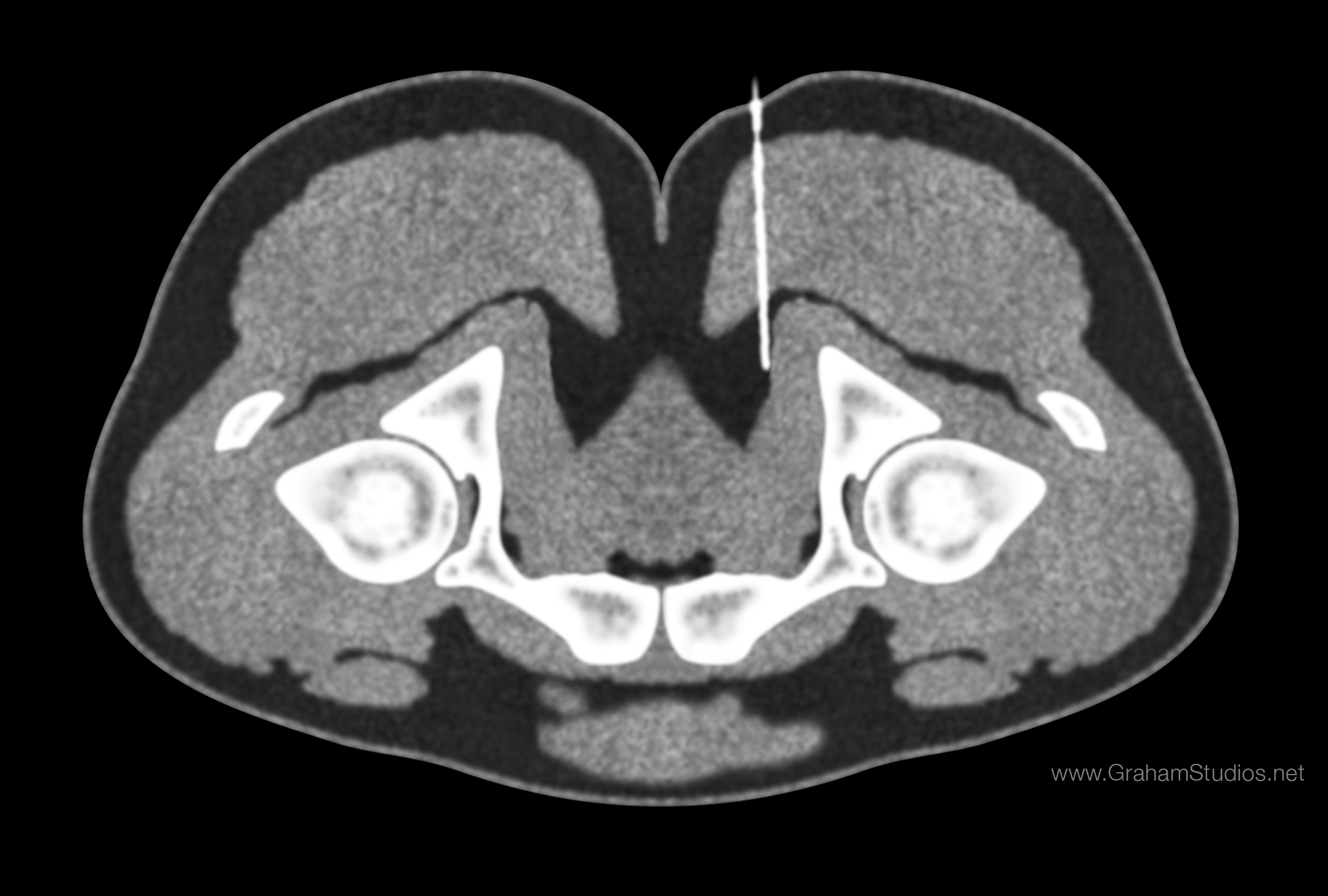
The pudendal nerve is difficult to visualize on routine CT or MR imaging, however under CT guidance, a needle may be placed adjacent to the pudendal neurovascular bundle. The ischial spine, an easily identifiable structure on CT, is used as the level of injection. A spinal needle is advanced via the gluteal muscles and advanced within several millimeters of the ischial spine. Contrast (X-ray dye) is then injected, highlighting the nerve in the canal and allowing for confirmation of correct needle placement. The nerve may then be injected with cortisone and local anesthetic to confirm and also treat chronic pain of the external genitalia (known as vulvodynia in females), pelvic and anorectal pain.
Nerve latency testing
The time taken for a muscle supplied by the pudendal nerve to contract in response to an electrical stimulus applied to the sensory and motor fibers can be quantified. Increased conduction time (terminal motor latency) signifies damage to the nerve. 2 stimulating electrodes and 2 measuring electrodes are mounted on the examiner's gloved finger ("St Mark's electrode").
History
The term pudendal comes from Latin pudenda, meaning external genitals, derived from pudendum, meaning "parts to be ashamed of". The pudendal canal is also known by the eponymous term "Alcock's canal", after Benjamin Alcock, an Irish anatomist who documented the canal in 1836. Alcock documented the existence of the canal and pudendal nerve in a contribution about iliac arteries in Robert Bentley Todd's "The Cyclopaedia of Anatomy and Physiology".
Additional images
File:Gray829.png|The male pelvis, showing the pudendal nerve (centre right) File:Grant 1962 214.png|Schematic showing the structures innervated by the pudendal nerve File:Grant 1962 215.png|Diagram of the course of the pudendal nerve in the male pelvis
References
References
- (2013). "Grant's atlas of anatomy". Wolters Kluwer Health/Lippincott Williams & Wilkins.
- Standring S (editor in chief). (2004). "Gray's Anatomy: The Anatomical Basis of Clinical Practice". Elsevier.
- Moore, Keith L. Moore, Anne M.R. Agur; in collaboration with and with content provided by Arthur F. Dalley II; with the expertise of medical illustrator Valerie Oxorn and the developmental assistance of Marion E.. (2007). "Essential clinical anatomy". Lippincott Williams & Wilkins.
- Russell RM. (2006). "Examination of peripheral nerve injuries an anatomical approach". Thieme.
- (2013). "Varney's midwifery". Jones & Bartlett Publishers.
- Nayak, Soubhagya R.. (November 2006). "Unusual origin of dorsal nerve of penis and abnormal formation of pudendal nerve—Clinical significance". Annals of Anatomy - Anatomischer Anzeiger.
- Shafik, A. (1995). "Surgical anatomy of the pudendal nerve and its clinical implications". Clinical Anatomy.
- (January 2022). "Pudendal Nerve Entrapment Syndrome.". StatPearls.
- Neill, editor-in-chief, Jimmy D.. (2006). "Knobil and Neill's physiology of reproduction". Elsevier.
- Ort, Bruce Ian Bogart, Victoria. (2007). "Elsevier's integrated anatomy and embryology". Elsevier Saunders.
- Babayan, Mike B. Siroky, Robert D. Oates, Richard K.. (2004). "Handbook of urology diagnosis and therapy". Lippincott Williams & Wilkins.
- Guaderrama, Noelani M.. (October 2005). "Evidence for the Innervation of Pelvic Floor Muscles by the Pudendal Nerve". Obstetrics & Gynecology.
- (2007). "The ASCRS textbook of colon and rectal surgery". Springer.
- Drake, Richard L.. (2005). "Gray's anatomy for students". Elsevier/Churchill Livingstone.
- (June 2008). "The neural control of micturition". Nat. Rev. Neurosci..
- Penson, David F.. (2002). "Male Sexual Function: A Guide to Clinical Management". Annals of Internal Medicine.
- (11 August 2020). "Clinical Decision Making in Colorectal Surgery". Springer International Publishing.
- (2002). "Maternal, Neonatal, and Women's Health Nursing, Volume 1". [[Cengage Learning]].
- "Transvaginal Pudendal Nerve Block". WebMD LLC.
- (2016). "Peripheral Nerve Entrapments: Clinical Diagnosis and Management". Springer International Publishing.
- Mellion MB. (January 1991). "Common cycling injuries. Management and prevention". Sports Med.
- (April 2018). "Pudendal nerve injury impairs anorectal function and health related quality of life measures ≥2 years after 3D conformal radiotherapy for prostate cancer". Taylor & Francis Group.
- Calvillo O, Skaribas IM, Rockett C.. (2000). "Computed tomography-guided pudendal nerve block. A new diagnostic approach to long-term anoperineal pain: a report of two cases". Reg Anesth Pain Med.
- Hough DM, Wittenberg KH, Pawlina W, Maus TP, King BF, Vrtiska TJ, Farrell MA, Antolak SJ Jr.. (2003). "Chronic perineal pain caused by pudendal nerve entrapment: anatomy and CT-guided perineural injection technique". Am J Roentgenol.
- G.A. Santoro, A.P. Wieczorek, C.I. Bartram (editors). (2010). "Pelvic floor disorders imaging and multidisciplinary approach to management". Springer.
- Harper, Douglas. "Pudendum". Online Etymology Dictionary.
- Oelhafen, Kim. (September 2013). "Benjamin Alcock (1801–?) and his canal". Clinical Anatomy.
This article was imported from Wikipedia and is available under the Creative Commons Attribution-ShareAlike 4.0 License. Content has been adapted to SurfDoc format. Original contributors can be found on the article history page.
Ask Mako anything about Pudendal nerve — get instant answers, deeper analysis, and related topics.
Research with MakoFree with your Surf account
Create a free account to save articles, ask Mako questions, and organize your research.
Sign up freeThis content may have been generated or modified by AI. CloudSurf Software LLC is not responsible for the accuracy, completeness, or reliability of AI-generated content. Always verify important information from primary sources.
Report