From Surf Wiki (app.surf) — the open knowledge base
Pseudoxanthoma elasticum
Genetic disease in which elastic fibers in certain tissues harden

Genetic disease in which elastic fibers in certain tissues harden
| Field | Value |
|---|---|
| synonyms | Grönblad–Strandberg syndrome; Groenblad-Strandberg syndrome |
| image | Pseudoxanthoma_elasticum_3.JPG |
| caption | Pseudoxanthoma elasticum of the posterior lateral neck. Note the yellowish slightly raised bumps characteristic of this condition. |
Pseudoxanthoma elasticum (PXE) is a genetic disease that causes mineralization of elastic fibers in some tissues. The most common problems arise in the skin and eyes, and later in blood vessels in the form of premature atherosclerosis. PXE is caused by autosomal recessive mutations in the ABCC6 gene on the short arm of chromosome 16 (16p13.1).
Signs and symptoms
Dermatological manifestations
Usually, pseudoxanthoma elasticum affects the skin first, often in childhood or early adolescence. Small, yellowish papular lesions form and cutaneous laxity mainly affect the neck, axillae (armpits), groin, and flexural creases (the inside parts of the elbows and knees). Skin may become lax and redundant. Many individuals have "oblique mental creases" (horizontal grooves of the chin). The lesions are asymptomatic and have a stochastic pattern of growth and spread.
Ocular manifestations
Pseudoxanthoma elasticum (PXE) causes progressive, abnormal calcification of Bruch's membrane in the central retina, producing a characteristic pattern of fundus changes and multiple vision-threatening complications. Early disease shows peau d'orange, a speckled mid-peripheral pattern marking the transition between normal retina and the enlarging central zone of confluent calcification. As Bruch's membrane becomes increasingly brittle, angioid streaks—fractures radiating from the optic disc—form and enlarge with age, predisposing strongly to choroidal neovascularization (CNV). CNV commonly emerges in mid-adulthood, recurs, and can lead to hemorrhage, scarring, and permanent loss of central vision. Independent of CNV, chronic dysfunction of the calcified membrane drives macular atrophy, causing enlarging scotomas and declining acuity.
Pre-clinical functional impairment includes delayed dark adaptation, likely from hindered nutrient and retinoid transport across the mineralized Bruch's membrane. Imaging also reveals choriocapillaris flow loss and reduced choroidal perfusion beneath calcified areas, likely another secondary alteration due to the mineralized Bruch's membrane.
PXE is also associated with a high prevalence of optic disc drusen, which correlate with disease severity and contribute to visual field defects and progressive inner retinal thinning. Finally, PXE eyes exhibit reduced ocular compliance, showing more prolonged intraocular pressure spikes after intravitreal injections—an important consideration when treating their frequent CNV.
Cardiovascular manifestations
PXE is caused by mineralization in connective tissues in mainly the skin, eyes, and blood vessels. As a result of mineralized buildup in the vascular wall, patients may be at a greater risk for intermittent claudication, a condition in which cramping pain in the leg is induced by exercise, and peripheral artery disease. Occlusions in cerebral arteries can lead to reduced or blocked blood flow, resulting in serious complications including transient ischemic attack (TIA) and stroke. Similarly, blockages in the coronary circulatory system, also called coronary artery disease may develop, leading to angina and myocardial infarction (heart attack). Cerebral ischemia in PXE is caused by small vessel occlusive disease.
Other manifestations
Although not much is known about potential gastroenterological manifestations, gastrointestinal bleeding is a rare symptom and usually involved bleeding from the stomach.
Other rare neurological complications may include intracranial aneurysms, subarachnoid and intracerebral hemorrhages.
Genetics
80% of clinical cases of pseudoxanthoma elasticum have detectable mutations in the ABCC6 gene. Mutations in almost all parts of the gene have been described, of all types (missense, nonsense, splice alteration, insertion, small deletion or large deletion). Although there have been reports of autosomal dominant inheritance, the inheritance is typically autosomal recessive (both parents need to be carriers, and there is a 25% chance that a child will inherit both abnormal copies of the gene and therefore develop the condition).
Strong genetic linkage was found with mutations in the ABCC6 gene, which codes for the ABCC6 protein, which is a membrane transporter from the large ATP-binding cassette transporter family. The protein is expressed in most organs, but mainly in the liver and kidney. ABCC6 mediates ATP release in the liver. This is the main source of circulating pyrophosphate (PPi), and individuals affected by PXE have strongly reduced plasma PPi levels, explaining their mineralization disorder. One study suggested that mutations causing total absence of an ABCC6 protein caused a more severe disease, but this could not be confirmed in a subsequent case series. Given the variations in age of onset and severity it is likely that other unknown risk factors (genetic, environmental, and lifestyle) may be involved.
Premature atherosclerosis is also associated with mutations in the ABCC6 gene, even in those without PXE. A syndrome almost indistinguishable from hereditary PXE has been described in patients with hemoglobinopathies (sickle-cell disease and thalassemia) through a poorly understood mechanism. Mutations in ABCC6 can also cause generalized arterial calcification of infancy. In some cases of PXE, mutations in ABCC6 cannot be found, and other genes such as ENPP1 may be implicated.
Pathophysiology
In PXE, there is mineralization (accumulation of calcium and other minerals) and fragmentation of the elastin-containing fibers in connective tissue, but primarily in the midlaminar layer of the dermis, Bruch's membrane and the midsized arteries. Recent studies have confirmed that PXE is a metabolic disease, and that its features arise because metabolites of vitamin K cannot reach peripheral tissues. Low levels of PPi cause mineralization in peripheral tissues.
Diagnosis
The diagnostic criteria for PXE are the typical skin biopsy appearance and the presence of angioid streaks in the retina. Criteria were established by consensus of clinicians and researchers at the 2010 biennial research meeting of the PXE Research Consortium. and confirmed at the 2014 meeting These consensus criteria state that definitive PXE is characterized by two pathogenic mutations in the ABCC6 or ocular findings – angioid streaks 1 DD or peau d'orange in an individual
- Characteristic pseudoxanthomatous papules and plaques on the neck or flexural creases.
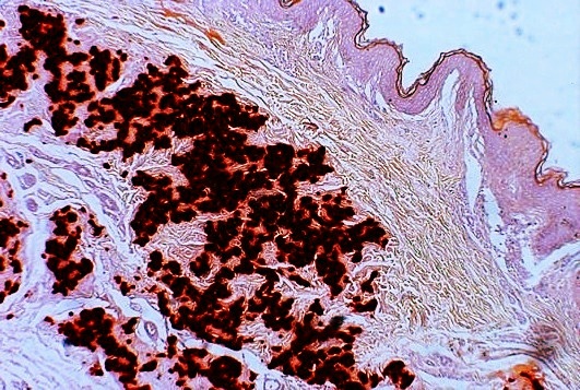
- Diagnostic histopathological changes in lesional skin: Calcified elastic fibers in the mid and lower dermis, confirmed by positive calcium stain
Treatment
There is no confirmed treatment that directly interferes with the disease process.
Cosmetic surgery to remove excessive skin has been used to improve aesthetic appearance in PXE patients but because of the non-life-threatening nature of these symptoms, should be used with caution.
One of the most critical symptom of PXE is choroidal neovascularization which can lead to deterioration of central vision. Photodynamic therapy has been used as a treatment, but this has been replaced with endothelial growth factor (VEGF) inhibitors (such as bevacizumab, ranibizumab, and aflibercept) with efficacy similar to their use in treatment of age-related macular degeneration.
To limit cardiovascular symptoms, reduction of cardiovascular risk factors through lifestyle changes is recommended. Generally clinicians recommend avoidance of non-steroidal anti-inflammatory drugs (NSAIDS) that increase bleeding risk, such as aspirin, and ibuprofen to prevent eye and gastrointestinal bleeding.
Formerly, dietary restriction of calcium was tried with no benefit, and in fact accelerated mineralization in mice. There are a number of potential treatments that are currently being tested or have just undergone testing including magnesium, etidronate, PPi, and tissue-nonspecific alkaline phosphatase inhibitors.
Given that ABCC6 heterozygous mutations result in few symptoms of PXE, this disease is a candidate for gene therapy. Some initial proof-of-principle experiments have been done in mice that have relieved some of symptoms of PXE, but as with all gene therapy treatments, there are many hurdles that must be overcome including insuring that the treatment will be long-lasting and reducing the risk of insertional mutagenesis and severe immune reactions.
Epidemiology
The reported prevalence of pseudoxanthoma elasticum is about 1:25,000. Females are twice as likely to be affected as males. The disease occurs in all ethnicities, but Afrikaners are more likely to have PXE as a result of a founder effect (i.e., higher prevalence in the small group of people from whom Afrikaners descend).
History
The first description of PXE that distinguished it from other xanthoma conditions was by Dr Ferdinand-Jean Darrier in 1896. The eponym "Grönblad-Strandberg syndrome" is used in older literature, after two physicians who made further discoveries in the disease manifestations.
PXE has the distinction of being the only disease for which a layperson is the discover of the mutated gene. The ABCC6 gene mutation was discovered simultaneously by four research teams, all of which published at the same time. The principal investigators were (in order of the date of publication): Jouni Uitto, Arthur Bergen, Charles Boyd, and Klaus Lindpainter. The gene was patented by Charles D. Boyd, Katalin Csiszar, Olivier LeSaux, Zsolt Urban, Sharon Terry, and assigned to PXE International by these co-inventors. Between the filing and 2013, when the Supreme Court of the United States declared that genes may not be patented. PXE International freely licensed the gene to any lab for clinical testing and research. PXE International continues to hold and maintain other patents (diagnosis and treatment patents).
PXE International, a support organization, was founded in 1995, by Patrick and Sharon Terry, following the diagnosis of their two children. It has a registry of 4,600 affected individuals.
Images
Image:Pseudoxanthoma_elasticum_1.JPG|Pseudoxanthoma elasticum of the posterior lateral neck. Image:Pseudoxanthoma_elasticum_2.JPG|Pseudoxanthoma elasticum of the left axillary fold.
References
References
- (2007). "Dermatology: 2-Volume Set". Mosby.
- (February 2019). "Pseudoxanthoma Elasticum as a Paradigm of Heritable Ectopic Mineralization Disorders: Pathomechanisms and Treatment Development". The American Journal of Pathology.
- (December 2005). "Pseudoxanthoma elasticum: a clinical, pathophysiological and genetic update including 11 novel ABCC6 mutations". Journal of Medical Genetics.
- (2009). "Pseudoxanthoma elasticum: genetics, clinical manifestations and therapeutic approaches". Survey of Ophthalmology.
- (August 2015). "Pseudoxanthoma elasticum and skin: Clinical manifestations, histopathology, pathomechanism, perspectives of treatment". Intractable & Rare Diseases Research.
- (May 2017). "Pseudoxanthoma elasticum". Orphanet Journal of Rare Diseases.
- (September 2009). "Manifestations of pseudoxanthoma elasticum in childhood". The British Journal of Dermatology.
- (2003). "Connective Tissue and Its Heritable Disorders: Molecular, Genetic, and Medical Aspects". John Wiley & Sons.
- (April 2003). "Prominent mental (chin) crease: a new sign of pseudoxanthoma elasticum". Journal of the American Academy of Dermatology.
- (2015). "Pseudoxanthoma elasticum and skin: Clinical manifestations, histopathology, pathomechanism, perspectives of treatment". Intractable & Rare Diseases Research.
- (September 2024). "Pseudoxanthoma elasticum – Genetics, pathophysiology, and clinical presentation". Progress in Retinal and Eye Research.
- (March 2024). "Bruch's Membrane Calcification in Pseudoxanthoma Elasticum". Ophthalmology Science.
- (2025-11-06). "High-Resolution Optical Coherence Tomography Correlates of Peau d'Orange in Pseudoxanthoma Elasticum". JAMA Ophthalmology.
- (December 2020). "The Extent of Angioid Streaks Correlates With Macular Degeneration in Pseudoxanthoma Elasticum". American Journal of Ophthalmology.
- (September 2024). "Anti-VEGF Treatment for Secondary Neovascularization in Pseudoxanthoma Elasticum - Age of Onset, Treatment Frequency, and Visual Outcome". American Journal of Ophthalmology.
- (2016-06-27). "Frequency, Phenotypic Characteristics and Progression of Atrophy Associated With a Diseased Bruch's Membrane in Pseudoxanthoma Elasticum". Investigative Ophthalmology & Visual Science.
- (October 2020). "Impaired Dark Adaptation Associated with a Diseased Bruch Membrane in Pseudoxanthoma Elasticum". Retina.
- (2025-02-06). "Topography of Slowed Dark Adaptation in Pseudoxanthoma Elasticum: PROPXE Study Report 1". Investigative Ophthalmology & Visual Science.
- (2025-11-13). "Measurement Reliability and Functional Validity of Bruch's Membrane Calcification in Pseudoxanthoma Elasticum: PROPXE Study Report 2". Investigative Ophthalmology & Visual Science.
- (2023-02-21). "Choriocapillaris Flow Signal Impairment in Patients With Pseudoxanthoma Elasticum". Investigative Ophthalmology & Visual Science.
- (2024-06-10). "Optic Disc Drusen in Pseudoxanthoma Elasticum Are Associated with the Extent of Bruch's Membrane Calcification". Journal of Clinical Medicine.
- (2025-08-11). "Intraocular Pressure After Anti-Vascular Endothelial Growth Factor Injection in Eyes With a Mineralized Bruch's Membrane Caused by Pseudoxanthoma Elasticum". Investigative Ophthalmology & Visual Science.
- (November 2011). "Relationship between ankle brachial index and arterial remodeling in pseudoxanthoma elasticum". Journal of Vascular Surgery.
- (January 2017). "Prevalence and severity of arterial calcifications in pseudoxanthoma elasticum (PXE) compared to hospital controls. Novel insights into the vascular phenotype of PXE". Atherosclerosis.
- (February 2017). "Cerebral disease in a nationwide Dutch pseudoxanthoma elasticum cohort with a systematic review of the literature". Journal of the Neurological Sciences.
- (May 2000). "Pseudoxanthoma elasticum: mutations in the MRP6 gene encoding a transmembrane ATP-binding cassette (ABC) transporter". Proceedings of the National Academy of Sciences of the United States of America.
- (June 2000). "Mutations in ABCC6 cause pseudoxanthoma elasticum". Nature Genetics.
- (June 2000). "Mutations in a gene encoding an ABC transporter cause pseudoxanthoma elasticum". Nature Genetics.
- (2000). "Mutations of the gene encoding the transmembrane transporter protein ABC-C6 cause pseudoxanthoma elasticum". Journal of Molecular Medicine.
- (September 2014). "ABCC6-mediated ATP secretion by the liver is the main source of the mineralization inhibitor inorganic pyrophosphate in the systemic circulation-brief report". Arteriosclerosis, Thrombosis, and Vascular Biology.
- (June 2005). "Novel mutations in the ABCC6 gene of German patients with pseudoxanthoma elasticum". Human Biology.
- (October 2007). "Mutation detection in the ABCC6 gene and genotype-phenotype analysis in a large international case series affected by pseudoxanthoma elasticum". Journal of Medical Genetics.
- (August 2002). "Frequent mutation in the ABCC6 gene (R1141X) is associated with a strong increase in the prevalence of coronary artery disease". Circulation.
- (March 2014). "Mutations in the ABCC6 gene as a cause of generalized arterial calcification of infancy: genotypic overlap with pseudoxanthoma elasticum". The Journal of Investigative Dermatology.
- (May 2015). "Genetic heterogeneity of pseudoxanthoma elasticum: the Chinese signature profile of ABCC6 and ENPP1 mutations". The Journal of Investigative Dermatology.
- (2003). "Extracutaneous ultrastructural alterations in pseudoxanthoma elasticum". Ultrastructural Pathology.
- (January 2009). "Pseudoxanthoma elasticum: clinical phenotypes, molecular genetics and putative pathomechanisms". Experimental Dermatology.
- (2012). "Histopathology of Pseudoxanthoma Elasticum and Related Disorders: Histological Hallmarks and Diagnostic Clues". Scientifica.
- (July 2011). "Pseudoxanthoma elasticum: progress in diagnostics and research towards treatment: Summary of the 2010 PXE International Research Meeting". American Journal of Medical Genetics. Part A.
- (June 2014). "Visual Acuity in Pseudoxanthoma Elasticum". Expert Opinion on Orphan Drugs.
- (September 2012). "Restricting dietary magnesium accelerates ectopic connective tissue mineralization in a mouse model of pseudoxanthoma elasticum (Abcc6(-/-) )". Experimental Dermatology.
- (July 2019). "Magnesium supplementation in the treatment of pseudoxanthoma elasticum: A randomized trial". Journal of the American Academy of Dermatology.
- (March 2018). "Etidronate for Prevention of Ectopic Mineralization in Patients With Pseudoxanthoma Elasticum". Journal of the American College of Cardiology.
- (November 2017). "Oral administration of pyrophosphate inhibits connective tissue calcification". EMBO Molecular Medicine.
- (February 2019). "-/- Mouse Model of PXE but Not in the Enpp1 Mutant Mouse Models of GACI". The Journal of Investigative Dermatology.
- (October 2002). "Evidence for a founder effect for pseudoxanthoma elasticum in the Afrikaner population of South Africa". Human Genetics.
- (1896). "Pseudoxanthoma elasticum". Monatschr Prakt Dermatol.
- {{WhoNamedIt. synd. 1059
- Liptak, Adam. (2013-06-13). "Justices, 9-0, Bar Patenting Human Genes". The New York Times.
- "Methods and Composition for Diagnosing and Treating Pseudoxanthoma Elasticum and Related Conditions". United States Patent and Trademark Office.
- "Methods for Diagnosing Pseudoxanthoma Elasticum". United States Patent and Trademark Office.
- (2003). "Learning genetics". Health Affairs.
- (March 2016). "Life as a numerator: Putting the person in personal genomics". Applied & Translational Genomics.
- Terry, Sharon. (15 June 2017). "Science didn't understand my kids' rare disease until I decided to study it".
- (29 September 2017). "Sharon Terry: When Siblings Get A Rare Diagnosis, Can Their Parents Find the Cure? TED Radio Hour.".
This article was imported from Wikipedia and is available under the Creative Commons Attribution-ShareAlike 4.0 License. Content has been adapted to SurfDoc format. Original contributors can be found on the article history page.
Ask Mako anything about Pseudoxanthoma elasticum — get instant answers, deeper analysis, and related topics.
Research with MakoFree with your Surf account
Create a free account to save articles, ask Mako questions, and organize your research.
Sign up freeThis content may have been generated or modified by AI. CloudSurf Software LLC is not responsible for the accuracy, completeness, or reliability of AI-generated content. Always verify important information from primary sources.
Report