From Surf Wiki (app.surf) — the open knowledge base
Pathogenic bacteria
Disease-causing bacteria
_and_YLLs_(B),_by_pathogen_and_GBD_super-region,_2019.jpg)
Disease-causing bacteria
| Field | Value |
|---|---|
| name | Pathogenic bacteria |
| image | Neisseria gonorrhoea in pus - Gram stain.jpg |
| caption | Neisseria gonorrhoeae (small red dots) in pus from a man with a urethral discharge (Gram stain) |
Pathogenic bacteria are bacteria that can cause disease. This article focuses on the bacteria that are pathogenic to humans. Most species of bacteria are harmless and many are beneficial but others can cause infectious diseases. The number of these pathogenic species in humans is estimated to be fewer than a hundred. By contrast, several thousand species are considered part of the gut flora, with a few hundred species present in each individual human's digestive tract.
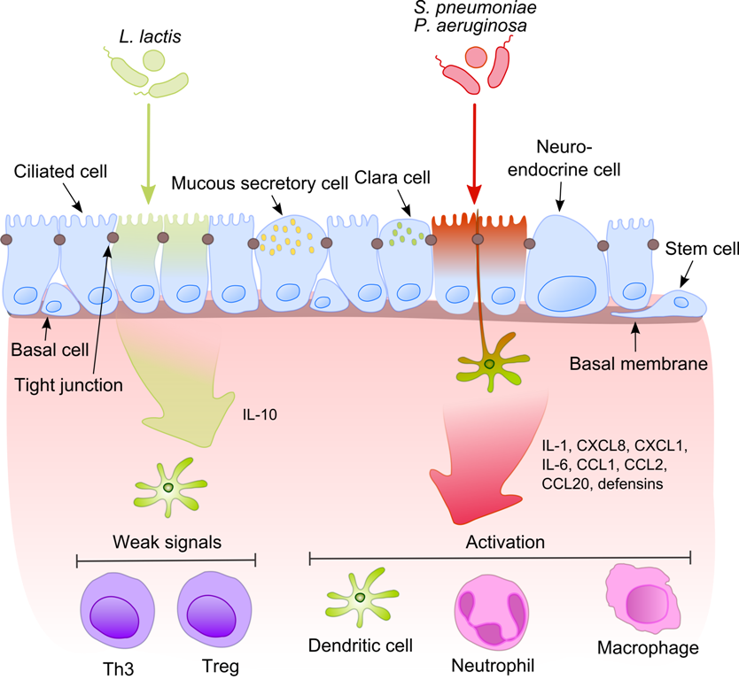
The body is continually exposed to many species of bacteria, including beneficial commensals, which grow on the skin and mucous membranes, and saprophytes, which grow mainly in the soil and in decaying matter. The blood and tissue fluids contain nutrients sufficient to sustain the growth of many bacteria. The body has defence mechanisms that enable it to resist microbial invasion of its tissues and give it a natural immunity or innate resistance against many microorganisms.
Pathogenic bacteria are specially adapted and endowed with mechanisms for overcoming the normal body defences, and can invade parts of the body, such as the blood, where bacteria are not normally found. Some pathogens invade only the surface epithelium, skin or mucous membrane, but many travel more deeply, spreading through the tissues and disseminating by the lymphatic and blood streams. In some rare cases a pathogenic microbe can infect an entirely healthy person, but infection usually occurs only if the body's defence mechanisms are damaged by some local trauma or an underlying debilitating disease, such as wounding, intoxication, chilling, fatigue, and malnutrition. In many cases, it is important to differentiate infection and colonization, which is when the bacteria are causing little or no harm.
Caused by Mycobacterium tuberculosis bacteria, one of the diseases with the highest disease burden is tuberculosis, which killed 1.4 million people in 2019, mostly in sub-Saharan Africa. Pathogenic bacteria contribute to other globally important diseases, such as pneumonia, which can be caused by bacteria such as Staphylococcus, * Streptococcus* and Pseudomonas, and foodborne illnesses, which can be caused by bacteria such as Shigella, Campylobacter, and Salmonella. Pathogenic bacteria also cause infections such as tetanus, typhoid fever, diphtheria, syphilis, and leprosy.
Pathogenic bacteria are also the cause of high infant mortality rates in developing countries. A GBD study estimated the global death rates from (33) bacterial pathogens, finding such infections contributed to one in 8 deaths (or ~7.7 million deaths), which could make it the second largest cause of death globally in 2019.
Most pathogenic bacteria can be grown in cultures and identified by Gram stain and other methods. Bacteria grown in this way are often tested to find which antibiotics will be an effective treatment for the infection. For hitherto unknown pathogens, Koch's postulates are the standard to establish a causative relationship between a microbe and a disease.
Diseases
Each species has specific effect and causes symptoms in people who are infected. Some people who are infected with a pathogenic bacteria do not have symptoms. Immunocompromised individuals are more susceptible to pathogenic bacteria.
Pathogenic susceptibility
Some pathogenic bacteria cause disease under certain conditions, such as entry through the skin via a cut, through sexual activity or through compromised immune function.
Some species of Streptococcus and Staphylococcus are part of the normal skin microbiota and typically reside on healthy skin or in the nasopharyngeal region. Yet these species can potentially initiate skin infections. Streptococcal infections include sepsis, pneumonia, and meningitis. These infections can become serious creating a systemic inflammatory response resulting in massive vasodilation, shock, and death.
Other bacteria are opportunistic pathogens and cause disease mainly in people with immunosuppression or cystic fibrosis. Examples of these opportunistic pathogens include Pseudomonas aeruginosa, Burkholderia cenocepacia, and Mycobacterium avium.
Intracellular
Obligate intracellular parasites (e.g. Chlamydophila, Ehrlichia, Rickettsia) are only able to grow and replicate inside other cells. Infections due to obligate intracellular bacteria may be asymptomatic, requiring an incubation period. Examples of obligate intracellular bacteria include Rickettsia prowazekii (typhus) and Rickettsia rickettsii, (Rocky Mountain spotted fever).
Chlamydia are intracellular parasites. These pathogens can cause pneumonia or urinary tract infection and may be involved in coronary heart disease.
Other groups of intracellular bacterial pathogens include Salmonella, Neisseria, Brucella, Mycobacterium, Nocardia, Listeria, Francisella, Legionella, and Yersinia pestis. These can exist intracellularly, but can exist outside host cells.
Infections in specific tissue
Bacterial pathogens often cause infection in specific areas of the body. Others are generalists.
- Bacterial vaginosis is a condition of the vaginal microbiota in which an excessive growth of Gardnerella vaginalis and other mostly anaerobic bacteria displace the beneficial Lactobacilli species that maintain healthy vaginal microbial populations.
- Bacterial meningitis is a bacterial inflammation of the meninges, which are the protective membranes covering the brain and spinal cord.
- Bacterial pneumonia is a bacterial infection of the lungs.
- Urinary tract infection is predominantly caused by bacteria. Symptoms include the strong and frequent sensation or urge to urinate, pain during urination, and urine that is cloudy. The most frequent cause is Escherichia coli. Urine is typically sterile but contains a variety of salts and waste products. Bacteria can ascend into the bladder or kidney and causing cystitis and nephritis.
- Bacterial gastroenteritis is caused by enteric, pathogenic bacteria. These pathogenic species are usually distinct from the usually harmless bacteria of the normal gut flora. But a different strain of the same species may be pathogenic. The distinction is sometimes difficult as in the case of Escherichia.
- Bacterial skin infections include:
- Impetigo is a highly contagious bacterial skin infection commonly seen in children. It is caused by Staphylococcus aureus, and Streptococcus pyogenes.
- Erysipelas is an acute streptococcus bacterial infection of the deeper skin layers that spreads via with lymphatic system.
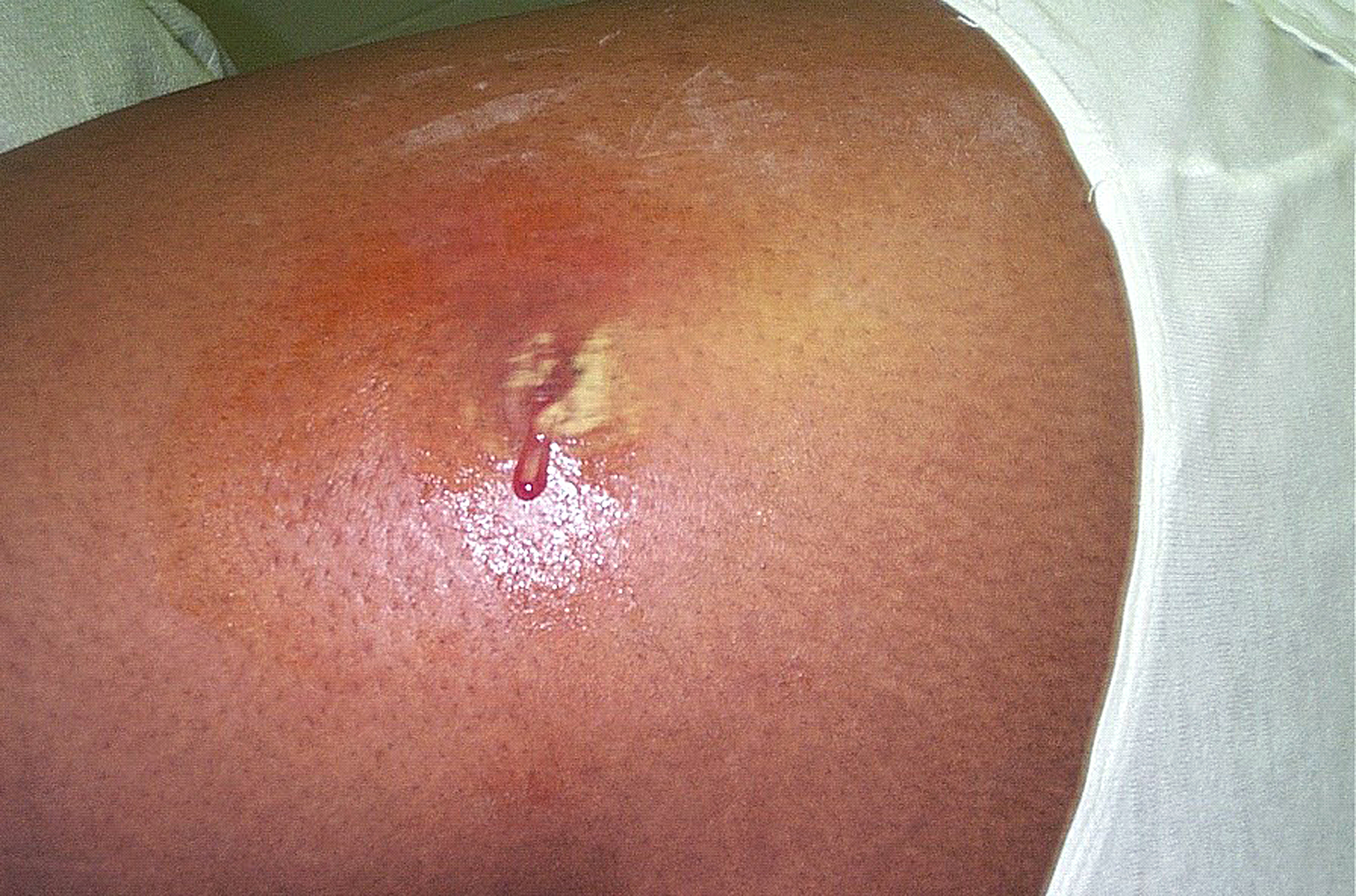
- Cellulitis is a diffuse inflammation of connective tissue with severe inflammation of dermal and subcutaneous layers of the skin. Cellulitis can be caused by normal skin flora or by contagious contact, and usually occurs through open skin, cuts, blisters, cracks in the skin, insect bites, animal bites, burns, surgical wounds, intravenous drug injection, or sites of intravenous catheter insertion. In most cases it is the skin on the face or lower legs that is affected, though cellulitis can occur in other tissues.
Mechanisms of damage
The symptoms of disease appear as pathogenic bacteria damage host tissues or interfere with their function. The bacteria can damage host cells directly or indirectly by provoking an immune response that inadvertently damages host cells, or by releasing toxins.
Direct
Once pathogens attach to host cells, they can cause direct damage as the pathogens use the host cell for nutrients and produce waste products. For example, Streptococcus mutans, a component of dental plaque, metabolizes dietary sugar and produces acid as a waste product. The acid decalcifies the tooth surface to cause dental caries.
Toxin production

Endotoxins are the lipid portions of lipopolysaccharides that are part of the outer membrane of the cell wall of gram-negative bacteria. Endotoxins are released when the bacteria lyses, which is why after antibiotic treatment, symptoms can worsen at first as the bacteria are killed and they release their endotoxins. Exotoxins are secreted into the surrounding medium or released when the bacteria die and the cell wall breaks apart.
Indirect
An excessive or inappropriate immune response triggered by an infection may damage host cells.
Survival in host
Nutrients
Iron is required for humans, as well as the growth of most bacteria. To obtain free iron, some pathogens secrete proteins called siderophores, which take the iron away from iron-transport proteins by binding to the iron even more tightly. Once the iron-siderophore complex is formed, it is taken up by siderophore receptors on the bacterial surface and then that iron is brought into the bacterium.
Bacterial pathogens also require access to carbon and energy sources for growth. To avoid competition with host cells for glucose which is the main energy source used by human cells, many pathogens including the respiratory pathogen Haemophilus influenzae specialise in using other carbon sources such as lactate that are abundant in the human body
Identification

Typically identification is done by growing the organism in a wide range of cultures which can take up to 48 hours. The growth is then visually or genomically identified. The cultured organism is then subjected to various assays to observe reactions to help further identify species and strain.
Treatment
Main article: Antibiotics
Bacterial infections may be treated with antibiotics, which are classified as bacteriocidal if they kill bacteria or bacteriostatic if they just prevent bacterial growth. There are many types of antibiotics and each class inhibits a process that is different in the pathogen from that found in the host. For example, the antibiotics chloramphenicol and tetracyclin inhibit the bacterial ribosome but not the structurally different eukaryotic ribosome, so they exhibit selective toxicity. Antibiotics are used both in treating human disease and in intensive farming to promote animal growth. Both uses may be contributing to the rapid development of antibiotic resistance in bacterial populations. Phage therapy, using bacteriophages can also be used to treat certain bacterial infections.
Prevention
Infections can be prevented by antiseptic measures such as sterilizing the skin prior to piercing it with the needle of a syringe and by proper care of indwelling catheters. Surgical and dental instruments are also sterilized to prevent infection by bacteria. Disinfectants such as bleach are used to kill bacteria or other pathogens on surfaces to prevent contamination and further reduce the risk of infection. Bacteria in food are killed by cooking to temperatures above 73 °C (163 °F).
List of genera and microscopy features
Many genera contain pathogenic bacterial species. They often possess characteristics that help to classify and organize them into groups. The following is a partial listing.
| Genus | Species | Gram staining | Shape | Oxygen requirement | Intra/Extracellular | |||||||||||||
|---|---|---|---|---|---|---|---|---|---|---|---|---|---|---|---|---|---|---|
| author1=Fisher, Bruce | author2=Harvey, Richard P. | author3=Champe, Pamela C. | title=Lippincott's Illustrated Reviews: Microbiology (Lippincott's Illustrated Reviews Series) | publisher=Lippincott Williams & Wilkins | location=Hagerstown, MD | year= 2007 | pages=332–353 | isbn=978-0-7817-8215-9 }} | Positive | Rods | Facultative anaerobic | Extracellular | ||||||
| Bartonella | Negative | Rods | Aerobic | Facultative intracellular | ||||||||||||||
| Bordetella | Negative | Small coccobacilli | Aerobic | Extracellular | ||||||||||||||
| Borrelia | Negative, stains poorly | Spirochete | Anaerobic | Extracellular | ||||||||||||||
| Brucella | Negative | Coccobacilli | Aerobic | Intracellular | ||||||||||||||
| Campylobacter | Negative | vauthors=Epps SV, Harvey RB, Hume ME, Phillips TD, Anderson RC, Nisbet DJ | title=Foodborne Campylobacter: infections, metabolism, pathogenesis and reservoirs | journal=International Journal of Environmental Research and Public Health | volume=10 | issue=12 | pages=6292–304 | year=2013 | pmid=24287853 | pmc=3881114 | doi=10.3390/ijerph10126292 | doi-access=free }} | ||||||
| coccoid in older cultures | Microaerophilic | Extracellular | ||||||||||||||||
| Chlamydia and Chlamydophila | (not Gram-stained) | Small, round, ovoid | Facultative or strictly aerobic | Obligate intracellular | ||||||||||||||
| Clostridium | Positive | Large, blunt-ended rods | Obligate anaerobic | Extracellular | ||||||||||||||
| Corynebacterium | Positive (unevenly) | Rods | Mostly facultative anaerobic | Extracellular | ||||||||||||||
| url=http://www.life.umd.edu/classroom/bsci424/LabMaterialsMethods/BSCI424Media.htm | title=BSCI424 Laboratory Media | access-date=2008-11-18 | first=David M. | last=Rollins | date=2000 | publisher=University of Maryland}} | Positive | Cocci | Facultative Anaerobic | Extracellular | ||||||||
| url=http://iws2.collin.edu/dcain/CCCCD%20Micro/macconkeyagar.htm | title=MacConkey Agar (CCCCD Microbiology | first=Donna | last=Cain | date=January 14, 2015 | publisher=Collin College | access-date=May 3, 2015 | archive-url=https://web.archive.org/web/20150426154407/http://iws2.collin.edu/dcain/CCCCD%20Micro/macconkeyagar.htm | archive-date=April 26, 2015}} | Negative | Rods | Facultative anaerobic | Extracellular or Intracellular | ||||||
| Francisella | Negative | Coccobacillus | Strictly aerobic | Facultative intracellular | ||||||||||||||
| Haemophilus | Negative | Coccobacilli to long and slender filaments | Facultative anaerobic 5 – 10% CO2 | Extracellular | ||||||||||||||
| Helicobacter | Negative | Spiral rod | Microaerophile | Extracellular | ||||||||||||||
| Legionella | Negative, stains poorly | Cocobacilli | Aerobic | Facultative intracellular | ||||||||||||||
| vauthors=Johnson RC, Harris VG | title=Differentiation of Pathogenic and Saprophytic Leptospires I. Growth at Low Temperatures | journal=J. Bacteriol. | volume=94 | issue=1 | pages=27–31 | year=1967 | doi=10.1128/JB.94.1.27-31.1967 | pmid=6027998 | pmc=251866}} | Negative, stains poorly | Spirochete | Strictly aerobic | Extracellular | |||||
| Listeria | Positive, darkly | Slender, short rods | Facultative Anaerobic | Facultative intracellular | ||||||||||||||
| Mycobacterium | (none) | Long, slender rods | Aerobic | Intracellular | ||||||||||||||
| Mycoplasma | (none) | Indistinct 'fried egg' appearance, no cell wall | Mostly facultative anaerobic; M. pneumoniae strictly aerobic | Extracellular | ||||||||||||||
| url=http://webmedia.unmc.edu/alliedhealth/CLS/CLS418%2008/Thayer%20Martin%20Agar%20Procedure%2008.pdf | title=Thayer Martin Agar (Modified) Procedure | publisher=University of Nebraska Medical Center, Clinical Laboratory Science Program | access-date=2015-05-03}} | Negative | Kidney bean-shaped | Aerobic | Gonococcus: facultative intracellular* | |||||||||||
| N. meningitidis*: extracellular | ||||||||||||||||||
| url=http://www.microbelibrary.org/component/resource/laboratory-test/2855-macconkey-agar-plates-protocols | title=MacConkey Agar Plates Protocols | publisher=American Society for Microbiology | year=2005 | first=Mary E. | last=Allen | archive-url=https://web.archive.org/web/20150507033028/http://www.microbelibrary.org/component/resource/laboratory-test/2855-macconkey-agar-plates-protocols | archive-date=2015-05-07}} Created: 30 September 2005. Last update: 01 April 2013 | Negative | Rods | Obligate aerobic | Extracellular | |||||||
| Rickettsia | Negative, stains poorly | Small, rod-like coccobacillary | Aerobic | Obligate intracellular | ||||||||||||||
| Salmonella | Negative | Rods | Facultative anaerobic | Facultative intracellular | ||||||||||||||
| url=http://www.austincc.edu/microbugz/hektoen_enteric_agar.php | title=Hektoen Enteric Agar | publisher=Austin Community College District | access-date=2015-05-03 | archive-date=2015-04-29 | archive-url=https://web.archive.org/web/20150429141248/http://www.austincc.edu/microbugz/hektoen_enteric_agar.php}} | Negative | Rods | Facultative anaerobic | Extracellular | |||||||||
| Staphylococcus | Positive, darkly | Round cocci | Facultative anaerobic | Extracellular, facultative intracellular | ||||||||||||||
| Streptococcus | Positive | Ovoid to spherical | Facultative anaerobic | Extracellular | ||||||||||||||
| Treponema | Negative, stains poorly | Spirochete | Aerobic | Extracellular | ||||||||||||||
| Ureaplasma | Stains poorly | Indistinct, 'fried egg' appearance, no cell wall | Anaerobic | Extracellular | ||||||||||||||
| last1=Pfeffer | first1=C. | last2=Oliver | first2=J.D. | year=2003 | title= A comparison of thiosulphate-citrate-bile salts-sucrose (TCBS) agar and thiosulphate-chloride-iodide (TCI) agar for the isolation of Vibrio species from estuarine environments | journal=Letters in Applied Microbiology | volume=36 | pmid=12581373 | issue=3 | pages=150–151 | doi=10.1046/j.1472-765X.2003.01280.x | s2cid=34004290 | doi-access=free }} | Negative | Spiral with single polar flagellum | Facultative anaerobic | Extracellular | |
| url=http://www.health.ny.gov/guidance/oph/wadsworth/yersinia_pestis.pdf | title=Yersinia pestis | publisher=Wadsworth Center | date=2006}} | Negative, bipolarly | Small rods | Facultative anaerobe | Intracellular |
List of species and clinical characteristics
File:Overall age-standardised mortality rate per 100 000 population for 33 pathogens investigated, 2019.jpg|Overall age-standardised mortality rate per 100 000 population for 33 pathogens investigated, 2019 File:Global number of deaths (A) and YLLs (B), by pathogen and infectious syndrome, 2019.jpg|Global number of deaths (A) and YLLs (B), by pathogen and infectious syndrome, 2019 File:Global number of deaths, by pathogen, age, and sex groups, 2019.jpg|Global number of deaths, by pathogen, age, and sex groups, 2019 This is description of the more common genera and species presented with their clinical characteristics and treatments.
| Species | Transmission | Diseases | Treatment | Prevention | ||||||||||
|---|---|---|---|---|---|---|---|---|---|---|---|---|---|---|
| Actinomyces israelii | Oral flora | last=Brook | first=I | title=Actinomycosis: diagnosis and management. | journal=Southern Medical Journal | date=Oct 2008 | volume=101 | issue=10 | pages=1019–23 | pmid=18791528 | doi=10.1097/SMJ.0b013e3181864c1f | s2cid=19554893}} or gastrointestinal tract. | Prolonged penicillin G and drainage | |
| Bacillus anthracis | url=http://emergency.cdc.gov/agent/anthrax/faq/treatment.asp | title=CDC Anthrax Q & A: Treatment | access-date=4 April 2011 | archive-url=https://web.archive.org/web/20110505191339/http://emergency.cdc.gov/agent/anthrax/faq/treatment.asp | archive-date=5 May 2011}} | |||||||||
| Bacteroides fragilis | Gut flora | Abscesses in gastrointestinal tract, pelvic cavity and lungs | metronidazole | Wound care | ||||||||||
| Bordetella pertussis | Macrolides such as erythromycin, before paroxysmal stage | |||||||||||||
| Borrelia | B. burgdorferi | |||||||||||||
| last1=Barbour | first1=Alan G. | editor1-last=Kasper | editor1-first=Dennis L. | editor2-last=Fauci | editor2-first=Anthony S. | title=Harrison's Infectious Diseases | date=2017 | publisher=McGraw Hill Education | location=New York | isbn=978-1-259-83597-1 | pages=678–687 | edition=3rd | chapter=Relapsing Fever}} | |
| Pediculus humanus corporis body louse (B. recurrentis only) and Ornithodoros soft ticks | Relapsing fever | vauthors=Cutler SJ | title=Relapsing Fever Borreliae: A Global Review | journal=Clinics in Laboratory Medicine | volume=35 | issue=4 | pages=847–65 | year=2015 | pmid=26593261 | doi=10.1016/j.cll.2015.07.001 }} | Avoid areas where ticks are found | |||
| Brucella | B. abortus | |||||||||||||
| Campylobacter jejuni | ||||||||||||||
| Chlamydia | C. pneumoniae | Atypical pneumonia | ||||||||||||
| C. trachomatis | ||||||||||||||
| Chlamydophila psittaci | Inhalation of dust with secretions or feces from birds (e.g. parrots) | Psittacosis, mainly atypical pneumonia | - | |||||||||||
| Clostridium | C. botulinum | canned]] food, smoked fish and honey | ||||||||||||
| C. difficile | Fecal bacteriotherapy | |||||||||||||
| C. perfringens | Gas gangrene: | Appropriate food handling | ||||||||||||
| C. tetani | ||||||||||||||
| Corynebacterium diphtheriae | ||||||||||||||
| Ehrlichia | E. canis | |||||||||||||
| Dog tick | muscle aches]], and fatigue | |||||||||||||
| Enterococcus | E. faecalis | |||||||||||||
| Escherichia | E. coli (generally) | UTI: | ||||||||||||
| Enterotoxigenic E. coli (ETEC) | ||||||||||||||
| Enteropathogenic E. coli | ||||||||||||||
| Enteroinvasive E.coli (EIEC) | ||||||||||||||
| Enterohemorrhagic (EHEC), including E. coli O157:H7 | ||||||||||||||
| Francisella tularensis | url=https://www.cdc.gov/tularemia/signssymptoms/ | title=Signs & Symptoms | website=Centers for Disease Control and Prevention | date=13 December 2018}} Page last reviewed: October 26, 2015 Can cause severe pneumonia. | ||||||||||
| Haemophilus influenzae | Meningitis: | |||||||||||||
| Helicobacter pylori | (No vaccine or preventive drug) | |||||||||||||
| Klebsiella pneumoniae | ||||||||||||||
| Legionella pneumophila | (no vaccine or preventive drug) | |||||||||||||
| Leptospira species | Vaccine not widely used | |||||||||||||
| Listeria monocytogenes | (no vaccine) | |||||||||||||
| Mycobacterium | M. leprae | Tuberculoid form: | ||||||||||||
| M. tuberculosis | ||||||||||||||
| Mycoplasma pneumoniae | ||||||||||||||
| Neisseria | N. gonorrhoeae | Uncomplicated gonorrhea: | ||||||||||||
| N. meningitidis | ||||||||||||||
| Pseudomonas aeruginosa | Opportunistic; Infects damaged tissues or people with immunodeficiency. | Pseudomonas infection: | (no vaccine) | |||||||||||
| Nocardia asteroides | In soil | Nocardiosis: Pneumonia, endocarditis, keratitis, neurological or lymphocutaneous infection | TMP/SMX | |||||||||||
| Rickettsia rickettsii | (no preventive drug or approved vaccine) | |||||||||||||
| Salmonella | S typhi | |||||||||||||
| Other Salmonella species | (No vaccine or preventive drug) | |||||||||||||
| Shigella | S. sonnei | |||||||||||||
| Staphylococcus | aureus | Coagulase-positive staphylococcal infections: | ||||||||||||
| epidermidis | Human flora in skin, anterior nares and mucous membranes | None | ||||||||||||
| saprophyticus | Part of normal vaginal flora | None | ||||||||||||
| Streptococcus | agalactiae | Human flora in vagina, urethral mucous membranes, rectum | ||||||||||||
| pneumoniae | ||||||||||||||
| pyogenes | No vaccine | |||||||||||||
| viridans | Oral flora, penetration through abrasions | Penicillin G | ||||||||||||
| Treponema pallidum subspecies pallidum | ||||||||||||||
| Vibrio cholerae | ||||||||||||||
| Yersinia pestis | Plague: |
Genetic transformation
Of the 59 species listed in the table with their clinical characteristics, 11 species (or 19%) are known to be capable of natural genetic transformation. Natural transformation is a bacterial adaptation for transferring DNA from one cell to another. This process includes the uptake of exogenous DNA from a donor cell by a recipient cell and its incorporation into the recipient cell's genome by recombination. Transformation appears to be an adaptation for repairing damage in the recipient cell's DNA. Among pathogenic bacteria, transformation capability likely serves as an adaptation that facilitates survival and infectivity. The pathogenic bacteria able to carry out natural genetic transformation (of those listed in the table) are Campylobacter jejuni, Enterococcus faecalis, Haemophilus influenzae, Helicobacter pylori, Klebsiella pneumoniae, Legionella pneumophila, Neisseria gonorrhoeae, Neisseria meningitidis, Staphylococcus aureus, Streptococcus pneumoniae and Vibrio cholerae.
Notes
References
References
- (2014). "Sherris Medical Microbiology". McGraw Hill Education.
- McFall-Ngai, Margaret. (2007-01-11). "Adaptive Immunity: Care for the community". Nature.
- (2022). "An expanded reference map of the human gut microbiome reveals hundreds of previously unknown species". Nature Communications.
- "Tuberculosis (TB)".
- (2013). "Risk of Early-Onset Neonatal Infection with Maternal Infection or Colonization: A Global Systematic Review and Meta-Analysis". PLOS Medicine.
- (23 November 2022). "Bacterial infections linked to 1 in 8 deaths in 2019". The Hill.
- (21 November 2022). "Global mortality associated with 33 bacterial pathogens in 2019: a systematic analysis for the Global Burden of Disease Study 2019". The Lancet.
- (February 2020). "Diagnosis of severe respiratory infections in immunocompromised patients". Intensive Care Medicine.
- "Streptococcal Infections - Infectious Diseases".
- Fish DN. (February 2002). "Optimal antimicrobial therapy for sepsis". Am J Health Syst Pharm.
- Heise E. (1982). "Diseases associated with immunosuppression". Environ Health Perspect.
- Saiman L. (2004). "Microbiology of early CF lung disease". Paediatr Respir Rev.
- (2004). "Chlamydia pneumoniae and atherosclerosis". Cell Microbiol.
- (August 2016). "Pathogenesis of Bacterial Vaginosis: Discussion of Current Hypotheses". The Journal of Infectious Diseases.
- "Urinary Tract Infections".
- Roxe DM. Urinalysis. In: Walker HK, Hall WD, Hurst JW, editors. Clinical Methods: The History, Physical, and Laboratory Examinations. 3rd edition. Boston: Butterworths; 1990. Chapter 191. Available from: https://www.ncbi.nlm.nih.gov/books/NBK302/
- (April 2018). "The challenge of urinary tract infections in renal transplant recipients". Transplant Infectious Disease.
- (19 October 2017). "Impetigo".
- Kumar, Vinay; Abbas, Abul K.; Fausto, Nelson; & Mitchell, Richard N. (2007). ''Robbins Basic Pathology'' (8th ed.). Saunders Elsevier. pp. 843 {{ISBN. 978-1-4160-2973-1
- {{DorlandsDict. three/000036667. erysipelas
- {{DorlandsDict. two/000019077. cellulitis
- (2012). "Medical Microbiology, a Guide to Microbial Infections: Pathogenesis, Immunity, Laboratory Investigation, and Control". Churchill Livingstone.
- (September 2017). "Bacterial toxins: Offensive, defensive, or something else altogether?". PLOS Pathogens.
- (2016). "Microbiology, an Introduction". Pearson Education.
- (2015). "Mims' Pathogenesis of Infectious Disease". Academic Press.
- Tortota, Gerard. (2013). "Microbiology an Introduction". Pearson.
- (2022-01-27). "Access to highly specialized growth substrates and production of epithelial immunomodulatory metabolites determine survival of Haemophilus influenzae in human airway epithelial cells". PLOS Pathogens.
- (2012). "Plant Cell Culture Protocols".
- (2004). "Ribosomal crystallography: initiation, peptide bond formation, and amino acid polymerization are hampered by antibiotics". Annu Rev Microbiol.
- Khachatourians GG. (November 1998). "Agricultural use of antibiotics and the evolution and transfer of antibiotic-resistant bacteria". CMAJ.
- (2012). "Phage Therapy: Concept to Cure". Frontiers in Microbiology.
- (2007). "Lippincott's Illustrated Reviews: Microbiology (Lippincott's Illustrated Reviews Series)". Lippincott Williams & Wilkins.
- (1988). "Comparison of modified Bordet-Gengou and modified Regan-Lowe media for the isolation of Bordetella pertussis and Bordetella parapertussis". J. Clin. Microbiol..
- (2007). "Lippincott's Illustrated Reviews: Microbiology (Lippincott's Illustrated Reviews Series)". Lippincott Williams & Wilkins.
- (2013). "Foodborne Campylobacter: infections, metabolism, pathogenesis and reservoirs". International Journal of Environmental Research and Public Health.
- Baron, Samuel. (1996). "Medical Microbiology". University of Texas Medical Branch at Galveston, Galveston, Texas.
- (2000). "Growth of Helicobacter pylori in various liquid and plating media". Lett. Appl. Microbiol..
- (1988). "Association of Ureaplasma urealyticum infection of the lower respiratory tract with chronic lung disease and death in very-low-birth-weight infants". Lancet.
- Brook, I. (Oct 2008). "Actinomycosis: diagnosis and management.". Southern Medical Journal.
- Mabeza, GF. (March 2003). "Pulmonary actinomycosis". European Respiratory Journal.
- Bowden GHW. (1996). "Actinomycosis ''in:'' Baron's Medical Microbiology". Univ of Texas Medical Branch.
- (2001). "Anthrax in animals". [[Food and Agriculture Organization]].
- "Bacteria Table". [[Creighton University School of Medicine]].
- "CDC Anthrax Q & A: Treatment".
- "FDA approves raxibacumab to treat inhalational anthrax".
- Itzhak Brook. (Jan 28, 2014). "Bacteroides Infection Follow-up". [[Medscape]].
- (2014). "Clinical practice. Lyme disease". The New England Journal of Medicine.
- (2015). "Clinical Manifestations and Treatment of Lyme Disease". Clinics in Laboratory Medicine.
- (2015). "Nervous System Lyme Disease". Clinics in Laboratory Medicine.
- (2017). "Harrison's Infectious Diseases". McGraw Hill Education.
- (2015). "Relapsing Fever Borreliae: A Global Review". Clinics in Laboratory Medicine.
- (May 2012). "Tetanus Epidemiology and Prevention of Vaccine-Preventable Diseases". Public Health Foundation.
- (20 January 2006). "Diphtheria vaccine". Wkly Epidemiol Rec.
- (2012-04-30). "ESCHERICHIA COLI". [[Public Health Agency of Canada]].
- (13 December 2018). "Signs & Symptoms".
- (2004). "Sherris Medical Microbiology". [[McGraw-Hill Education.
- (19 February 2021). "''Klebsiella pneumoniae'' in Healthcare Settings". [[Centers for Disease Control and Prevention]].
- Slack, A. (Jul 2010). "Leptospirosis.". Australian Family Physician.
- McBride, AJ. (Oct 2005). "Leptospirosis". Current Opinion in Infectious Diseases.
- (2017). "Harrison's Infectious Diseases". McGraw Hill Education.
- (January 2014). "Leprosy Fact sheet N°101". World Health Organization.
- (October 2015). "Tuberculosis Fact sheet N°104".
- [http://www.pasteur.fr/ip/easysite/go/03b-00002u-06f/press/press-releases/2009/vaccine-against-shigellosis-bacillary-dysentery-a-promising-clinical-trial Institut Pasteur Press Office - Vaccine against shigellosis (bacillary dysentery):a promising clinical trial] {{webarchive. link. (2009-02-25 15 January 2009. Retrieved on 27 February 2009)
- Levinson, W.. (2010). "Review of Medical Microbiology and Immunology".
- (November 2, 2015). "Syphilis - CDC Fact Sheet (Detailed)".
- (February 2008). "Reexamining syphilis: an update on epidemiology, clinical manifestations, and management". Annals of Pharmacotherapy.
- (2017). "Syphilis". Lancet.
- (2006). "Molecular and physiological insights into plague transmission, virulence and etiology". Microbes Infect..
- Wagle PM.. (1948). "Recent advances in the treatment of bubonic plague". Indian J Med Sci.
- Meyer KF.. (1950). "Modern therapy of plague". JAMA.
- (1992). "A decade of plague epidemiology and control in the Western Usambara mountains, north-east Tanzania". Acta Tropica.
- (September 2007). "Yersinia pestis CO92ΔyopH Is a Potent Live, Attenuated Plague Vaccine". Clin. Vaccine Immunol..
- Bernstein H, Bernstein C, Michod RE (2018). Sex in microbial pathogens. Infection, Genetics and Evolution volume 57, pages 8-25. https://doi.org/10.1016/j.meegid.2017.10.024
This article was imported from Wikipedia and is available under the Creative Commons Attribution-ShareAlike 4.0 License. Content has been adapted to SurfDoc format. Original contributors can be found on the article history page.
Ask Mako anything about Pathogenic bacteria — get instant answers, deeper analysis, and related topics.
Research with MakoFree with your Surf account
Create a free account to save articles, ask Mako questions, and organize your research.
Sign up freeThis content may have been generated or modified by AI. CloudSurf Software LLC is not responsible for the accuracy, completeness, or reliability of AI-generated content. Always verify important information from primary sources.
Report