From Surf Wiki (app.surf) — the open knowledge base
Neurofilament light polypeptide
Protein-coding gene in the species Homo sapiens
Protein-coding gene in the species Homo sapiens
Neurofilament light polypeptide is a protein that in humans is encoded by the NEFL gene.
Structure
Neurofilament light polypeptide is a member of the intermediate filament protein family. This protein family consists of over 50 human proteins divided into 5 major classes, the Class I and II keratins, Class III vimentin, GFAP, desmin and the others, the Class IV neurofilaments and the Class V nuclear lamins. There are four major neurofilament subunits, NF-L, NF-M, NF-H and α-internexin. These form heteropolymers which assemble to produce 10 nm neurofilaments which are only expressed in neurons where they are major structural proteins, particularly concentrated in large projection axons. The NF-L protein is encoded by the NEFL gene.
Function
These neurofilament heteropolymers assemble into the cytoskeleton of axons, where they provide structural support and help regulate axonal diameter and conduction velocity. Axons are particularly sensitive to mechanical and metabolic compromise and as a result axonal degeneration is a significant problem in many neurological disorders. Neurofilament light chain is a biomarker that can be measured with immunoassays in cerebrospinal fluid and plasma and reflects axonal damage in a wide variety of neurological disorders.
Measurement
NF-L antibodies employed in the most widely used NF-L assays are specific for cleaved forms of NF-L generated by proteolysis induced by cell death. Methods used in different studies for NfL measurement are sandwich enzyme-linked immunosorbent assay (ELISA), electrochemiluminescence, and high-sensitive single molecule array (SIMOA).
Clinical significance
The detection of neurofilament subunits in CSF and blood has become widely used as a biomarker of ongoing axonal compromise. It is a useful marker for disease monitoring in amyotrophic lateral sclerosis, multiple sclerosis, Alzheimer's disease, and more recently Huntington's disease. It is also a promising marker for follow-up of patients with brain tumors. Higher levels of blood or CSF NF-L have been associated with increased mortality, as would be expected as release of this protein reflects ongoing axonal loss.
It is associated with Charcot–Marie–Tooth disease 1F and 2E.
Neurofilament assembly

Neurofilament light polypeptide (NF-L) is a key structural component of the neuronal cytoskeleton, assembling into neurofilaments along with other intermediate filament proteins such as NF-M, NF-H, and α-internexin. These proteins form obligate heteropolymers that organize into 10 nm diameter filaments, which are selectively expressed in neurons and are particularly concentrated in axons[9]. Neurofilaments provide essential structural support, help maintain axonal diameter, and contribute to the efficient conduction of nerve impulses.
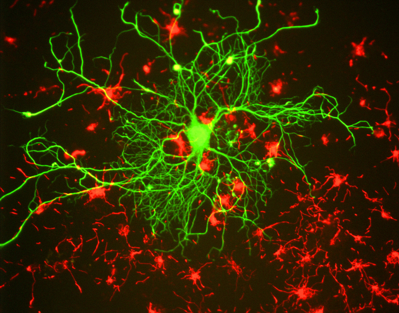
The localization and organization of NF-L in neurons can be visualized using immunohistochemical techniques. In tissue culture preparations of rat brain cells, antibodies specific to NF-L label large neurons prominently in green, revealing their extensive cytoskeletal architecture. In the same cultures, staining for α-internexin in red highlights surrounding neuronal stem cells, indicating the differential expression of these intermediate filament proteins during neural development and differentiation.
In histological sections of human brain tissue, NF-L can also be visualized using immunostaining. For example, in formalin-fixed and paraffin-embedded sections of the human cerebellum, an antibody specific to NF-L reveals its presence throughout various neuronal compartments[7]. The brown-stained antibody binding highlights the axonal processes of basket cells, the parallel fibers of granule cells, the perikarya of Purkinje cells, and other axonal elements. Counterstaining with a blue dye allows for the visualization of cell nuclei, delineating the granular layer on the left side of the section and the molecular layer on the right. These staining patterns underscore the widespread and structurally critical role of NF-L in both developing and mature neurons.
Interactions
Neurofilament light polypeptide has been shown to interact with:
- MAP2,
- Protein kinase N1, and
- TSC1.
References
References
- (July 2007). "Clinical and electrophysiological features in Charcot-Marie-Tooth disease with mutations in the NEFL gene". Archives of Neurology.
- "Entrez Gene: NEFL neurofilament, light polypeptide 68kDa".
- (October 2018). "Neurofilaments as biomarkers in neurological disorders". Nature Reviews. Neurology.
- (2023-03-02). "Uman-type neurofilament light antibodies are effective reagents for the imaging of neurodegeneration". Brain Communications.
- (September 2022). "Evaluation of cerebrospinal fluid neurofilament light chain levels in multiple sclerosis and non-demyelinating diseases of the central nervous system: clinical and biochemical perspective". Bosnian Journal of Basic Medical Sciences.
- (2016). "Neurofilaments as Biomarkers for Amyotrophic Lateral Sclerosis: A Systematic Review and Meta-Analysis". PLOS ONE.
- (2018). "Neurofilament light chain as a biological marker for multiple sclerosis: a meta-analysis study". Neuropsychiatric Disease and Treatment.
- (February 2019). "Biomarkers for Alzheimer's disease beyond amyloid and tau". Nature Medicine.
- (February 2019). "Serum neurofilament dynamics predicts neurodegeneration and clinical progression in presymptomatic Alzheimer's disease". Nature Medicine.
- (27 February 2017). "Tau or neurofilament light-Which is the more suitable biomarker for Huntington's disease?". PLOS ONE.
- (April 2022). "Evaluation of cerebrospinal fluid neurofilament light chain levels in multiple sclerosis and non-demyelinating diseases of the central nervous system: clinical and biochemical perspective". Bosnian Journal of Basic Medical Sciences.
- (February 2021). "A neuronal blood marker is associated with mortality in old age". Nature Aging.
- (April 2017). "Neurofilaments and Neurofilament Proteins in Health and Disease". Cold Spring Harbor Perspectives in Biology.
- (July 2012). "Neurofilaments at a glance". Journal of Cell Science.
- (2023). "Uman-type neurofilament light antibodies are effective reagents for the imaging of neurodegeneration". Brain Communications.
- (May 2021). "Interactions Between Purkinje Cells and Granule Cells Coordinate the Development of Functional Cerebellar Circuits". Neuroscience.
- (2025). "Neurofilaments". Synaptic Systems GmbH.
- (April 1991). "Interaction domains of neurofilament light chain and brain spectrin". The Biochemical Journal.
- (April 1996). "PKN associates and phosphorylates the head-rod domain of neurofilament protein". The Journal of Biological Chemistry.
- (November 2002). "The TSC1 tumor suppressor hamartin interacts with neurofilament-L and possibly functions as a novel integrator of the neuronal cytoskeleton". The Journal of Biological Chemistry.
This article was imported from Wikipedia and is available under the Creative Commons Attribution-ShareAlike 4.0 License. Content has been adapted to SurfDoc format. Original contributors can be found on the article history page.
Ask Mako anything about Neurofilament light polypeptide — get instant answers, deeper analysis, and related topics.
Research with MakoFree with your Surf account
Create a free account to save articles, ask Mako questions, and organize your research.
Sign up freeThis content may have been generated or modified by AI. CloudSurf Software LLC is not responsible for the accuracy, completeness, or reliability of AI-generated content. Always verify important information from primary sources.
Report