From Surf Wiki (app.surf) — the open knowledge base
Myosin
Family of motor proteins

Family of motor proteins
Myosins () are a family of motor proteins (though most often protein complexes) best known for their roles in muscle contraction and in a wide range of other motility processes in eukaryotes. They are ATP-dependent and responsible for actin-based motility.
The first myosin (M2) to be discovered was in 1864 by Wilhelm Kühne. Kühne had extracted a viscous protein from skeletal muscle that he held responsible for keeping the tension state in muscle. He called this protein myosin. The term has been extended to include a group of similar ATPases found in the cells of both striated muscle tissue and smooth muscle tissue.
Following the discovery in 1973 of enzymes with myosin-like function in Acanthamoeba castellanii, a global range of divergent myosin genes have been discovered throughout the realm of eukaryotes.
Although myosin was originally thought to be restricted to muscle cells (hence myo-(s) + -in), there is no single "myosin"; rather it is a very large superfamily of genes whose protein products share the basic properties of actin binding, ATP hydrolysis (ATPase enzyme activity), and force transduction. Virtually all eukaryotic cells contain myosin isoforms. Some isoforms have specialized functions in certain cell types (such as muscle), while other isoforms are ubiquitous. The structure and function of myosin is globally conserved across species, to the extent that rabbit muscle myosin II will bind to actin from an amoeba.
Structure and functions
Domains
Most myosin molecules are composed of a head, neck, and tail domain.
- The head domain binds the filamentous actin, and uses ATP hydrolysis to generate force and to "walk" along the filament towards the barbed (+) end (with the exception of myosin VI, which moves towards the pointed (-) end).
- the neck domain acts as a linker and as a lever arm for transducing force generated by the catalytic motor domain. The neck domain can also serve as a binding site for myosin light chains which are distinct proteins that form part of a macromolecular complex and generally have regulatory functions.
- The tail domain generally mediates interaction with cargo molecules and/or other myosin subunits. In some cases, the tail domain may play a role in regulating motor activity.
Power stroke
Main article: Muscle contraction
Multiple myosin II molecules generate force in skeletal muscle through a power stroke mechanism fuelled by the energy released from ATP hydrolysis. The power stroke occurs at the release of phosphate from the myosin molecule after the ATP hydrolysis while myosin is tightly bound to actin. The effect of this release is a conformational change in the molecule that pulls against the actin. The release of the ADP molecule leads to the so-called rigor state of myosin. The binding of a new ATP molecule will release myosin from actin. ATP hydrolysis within the myosin will cause it to bind to actin again to repeat the cycle. The combined effect of the myriad power strokes causes the muscle to contract.
Nomenclature, evolution, and the family tree

The wide variety of myosin genes found throughout the eukaryotic phyla were named according to different schemes as they were discovered. The nomenclature can therefore be somewhat confusing when attempting to compare the functions of myosin proteins within and between organisms.
Skeletal muscle myosin, the most conspicuous of the myosin superfamily due to its abundance in muscle fibers, was the first to be discovered. This protein makes up part of the sarcomere and forms macromolecular filaments composed of multiple myosin subunits. Similar filament-forming myosin proteins were found in cardiac muscle, smooth muscle, and nonmuscle cells. However, beginning in the 1970s, researchers began to discover new myosin genes in simple eukaryotes encoding proteins that acted as monomers and were therefore entitled Class I myosins. These new myosins were collectively termed "unconventional myosins" and have been found in many tissues other than muscle. These new superfamily members have been grouped according to phylogenetic relationships derived from a comparison of the amino acid sequences of their head domains, with each class being assigned a Roman numeral (see phylogenetic tree). The unconventional myosins also have divergent tail domains, suggesting unique functions. The now diverse array of myosins likely evolved from an ancestral precursor (see picture).
Analysis of the amino acid sequences of different myosins shows great variability among the tail domains, but strong conservation of head domain sequences. Presumably this is so the myosins may interact, via their tails, with a large number of different cargoes, while the goal in each case – to move along actin filaments – remains the same and therefore requires the same machinery in the motor. For example, the human genome contains over 40 different myosin genes.
These differences in shape also determine the speed at which myosins can move along actin filaments. The hydrolysis of ATP and the subsequent release of the phosphate group causes the "power stroke", in which the "lever arm" or "neck" region of the heavy chain is dragged forward. Since the power stroke always moves the lever arm by the same angle, the length of the lever arm determines the displacement of the cargo relative to the actin filament. A longer lever arm will cause the cargo to traverse a greater distance even though the lever arm undergoes the same angular displacement – just as a person with longer legs can move farther with each individual step. The velocity of a myosin motor depends upon the rate at which it passes through a complete kinetic cycle of ATP binding to the release of ADP.
Myosin classes
Myosin I
Myosin I, a ubiquitous cellular protein, functions as monomer and functions in vesicle transport. It has a step size of 10 nm and has been implicated as being responsible for the adaptation response of the stereocilia in the inner ear.
Myosin II

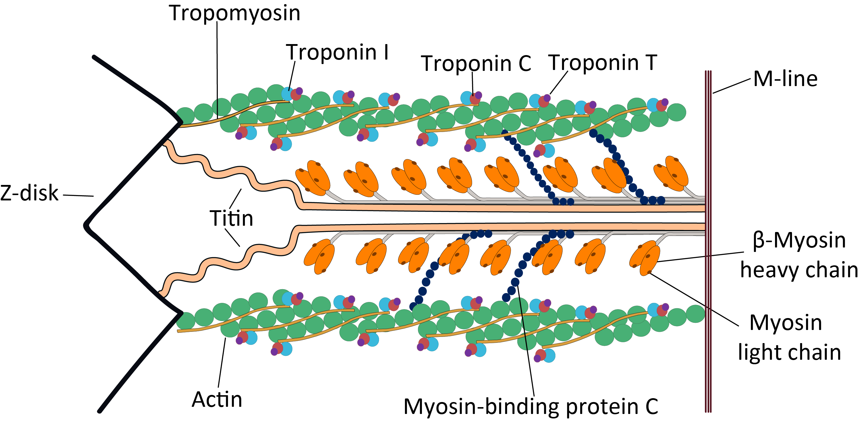
Myosin II (also known as conventional myosin) is the myosin type responsible for producing muscle contraction in muscle cells in most animal cell types. It is also found in non-muscle cells in contractile bundles called stress fibers.
- Myosin II contains two heavy chains, each about 2000 amino acids in length, which constitute the head and tail domains. Each of these heavy chains contains the N-terminal head domain, while the C-terminal tails take on a coiled-coil morphology, holding the two heavy chains together (imagine two snakes wrapped around each other, as in a caduceus). Thus, myosin II has two heads. The intermediate neck domain is the region creating the angle between the head and tail. In smooth muscle, a single gene (MYH11)) codes for the heavy chains myosin II, but splice variants of this gene result in four distinct isoforms.
- It also contains 4 myosin light chains (MLC), resulting in 2 per head, weighing 20 (MLC20) and 17 (MLC17) kDa. These bind the heavy chains in the "neck" region between the head and tail.
- [[File:MyosinII AU to rel.gif|thumb|Self-inhibition of Myosin II. The movie begins with Myosin II in the 10S conformation with a folded tail domain, the blocked head and free head. The movie schematically depicts tail unfolding and the resulting active 6S confirmation followed by tail folding back to the 10S conformation. The illustration is conceptual: transitory states and diffusive motions associated with folding/unfolding are not shown.]]The MLC20 is also known as the regulatory light chain and actively participates in muscle contraction.
- The MLC17 is also known as the essential light chain. Its exact function is unclear, but is believed to contribute to the structural stability of the myosin head along with MLC20. Two variants of MLC17 (MLC17a/b) exist as a result of alternative splicing at the MLC17 gene.
In muscle cells, the long coiled-coil tails of the individual myosin molecules can auto-inhibit active function in the 10S conformation or upon phosphorylation, change to the 6S conformation and join, forming the thick filaments of the sarcomere. The force-producing head domains stick out from the side of the thick filament, ready to walk along the adjacent actin-based thin filaments in response to the proper chemical signals and may be in either auto-inhibited or active conformation. The balance/transition between active and inactive states is subject to extensive chemical regulation.
Myosin III
Myosin III is a poorly understood member of the myosin family. It has been studied in vivo in the eyes of Drosophila, where it is thought to play a role in phototransduction. A human homologue gene for myosin III, MYO3A, has been uncovered through the Human Genome Project and is expressed in the retina and cochlea.[[File:Myosin V.png|thumbnail|right|Crystal structure of myosin V motor with essential light chain – nucleotide-free]]
Myosin IV
Myosin IV has a single IQ motif and a tail that lacks any coiled-coil forming sequence. It has homology similar to the tail domains of Myosin VII and XV.
Myosin V
Myosin V is an unconventional myosin motor, which is processive as a dimer and has a step size of 36 nm. It translocates (walks) along actin filaments traveling towards the barbed end (+ end) of the filaments. Myosin V is involved in the transport of cargo (e.g. RNA, vesicles, organelles, mitochondria) from the center of the cell to the periphery, but has been furthermore shown to act like a dynamic tether, retaining vesicles and organelles in the actin-rich periphery of cells. A recent single molecule in vitro reconstitution study on assembling actin filaments suggests that Myosin V travels farther on newly assembling (ADP-Pi rich) F-actin, while processive runlengths are shorter on older (ADP-rich) F-actin.

The Myosin V motor head can be subdivided into the following functional regions:
-
Nucleotide-binding site - These elements together coordinate di-valent metal cations (usually magnesium) and catalyze hydrolysis:
- Switch I - This contains a highly conserved SSR motif. Isomerizes in the presence of ATP.
- Switch II - This is the Kinase-GTPase version of the Walker B motif DxxG. Isomerizes in the presence of ATP.
- P-loop - This contains the Walker A motif GxxxxGK(S,T). This is the primary ATP binding site.
-
Transducer - The seven β-strands that underpin the motor head's structure.
-
U50 and L50 - The Upper (U50) and Lower (L50) domains are each around 50kDa. Their spatial separation forms a cleft critical for binding to actin and some regulatory compounds.
-
SH1 helix and Relay - These elements together provide an essential mechanism for coupling the enzymatic state of the motor domain to the powerstroke-producing region (converter domain, lever arm, and light chains).
-
Converter - This converts a change of conformation in the motor head to an angular displacement of the lever arm (in most cases reinforced with light chains).
Myosin VI

Myosin VI is an unconventional myosin motor, which is primarily processive as a dimer, but also acts as a nonprocessive monomer. It walks along actin filaments, travelling towards the pointed end (- end) of the filaments. Myosin VI is thought to transport endocytic vesicles into the cell.
Myosin VII
Myosin VII is an unconventional myosin with two FERM domains in the tail region. It has an extended lever arm consisting of five calmodulin binding IQ motifs followed by a single alpha helix (SAH) Myosin VII is required for phagocytosis in Dictyostelium discoideum, spermatogenesis in C. elegans and stereocilia formation in mice and zebrafish.
Myosin VIII
Myosin VIII is a plant-specific myosin linked to cell division; specifically, it is involved in regulating the flow of cytoplasm between cells and in the localization of vesicles to the phragmoplast.
Myosin IX
Myosin IX is a group of single-headed motor proteins. It was first shown to be minus-end directed, but a later study showed that it is plus-end directed. The movement mechanism for this myosin is poorly understood.
Myosin X
Myosin X is an unconventional myosin motor, which is functional as a dimer. The dimerization of myosin X is thought to be antiparallel. This behavior has not been observed in other myosins. In mammalian cells, the motor is found to localize to filopodia. Myosin X walks towards the barbed ends of filaments. Some research suggests it preferentially walks on bundles of actin, rather than single filaments. It is the first myosin motor found to exhibit this behavior.
Myosin XI
Myosin XI directs the movement of organelles such as plastids and mitochondria in plant cells. It is responsible for the light-directed movement of chloroplasts according to light intensity and the formation of stromules interconnecting different plastids. Myosin XI also plays a key role in polar root tip growth and is necessary for proper root hair elongation. A specific Myosin XI found in Nicotiana tabacum was discovered to be the fastest known processive molecular motor, moving at 7 μm/s in 35 nm steps along the actin filament.
Myosin XII
Myosin XIII
Myosin XIV
This myosin group has been found in the Apicomplexa phylum. The myosins localize to plasma membranes of the intracellular parasites and may then be involved in the cell invasion process.
This myosin is also found in the ciliated protozoan Tetrahymena thermophila. Known functions include: transporting phagosomes to the nucleus and perturbing the developmentally regulated elimination of the macronucleus during conjugation.
Myosin XV
Myosin XV is necessary for the development of the actin core structure of the non-motile stereocilia located in the inner ear. It is thought to be functional as a monomer.
Myosin XVI
Myosin XVII
Myosin XVIII
MYO18A A gene on chromosome 17q11.2 that encodes actin-based motor molecules with ATPase activity, which may be involved in maintaining stromal cell scaffolding required for maintaining intercellular contact.
Myosin XIX
Unconventional myosin XIX (Myo19) is a mitochondrial associated myosin motor.
Genes in humans
Note that not all of these genes are active.
- Class I: MYO1A, MYO1B, MYO1C, MYO1D, MYO1E, MYO1F, MYO1G, MYO1H
- Class II: MYH1, MYH2, MYH3, MYH4, MYH6, MYH7, MYH7B, MYH8, MYH9, MYH10, MYH11, MYH13, MYH14, MYH15, MYH16
- Class III: MYO3A, MYO3B
- Class V: MYO5A, MYO5B, MYO5C
- Class VI: MYO6
- Class VII: MYO7A, MYO7B
- Class IX: MYO9A, MYO9B
- Class X: MYO10
- Class XV: MYO15A, MYO15B
- Class XVI: MYO16
- Class XVIII: MYO18A, MYO18B
- Class XIX: MYO19
Myosin light chains are distinct and have their own properties. They are not considered "myosins" but are components of the macromolecular complexes that make up the functional myosin enzymes.
- Light chain: MYL1, MYL2, MYL3, MYL4, MYL5, MYL6, MYL6B, MYL7, MYL9, MYLIP, MYLK, MYLK2, MYLL1
Paramyosin
Paramyosin is a large, 93-115kDa muscle protein that has been described in a number of diverse invertebrate phyla. Invertebrate thick filaments are thought to be composed of an inner paramyosin core surrounded by myosin. The myosin interacts with actin, resulting in fibre contraction. Paramyosin is found in many different invertebrate species, for example, Brachiopoda, Sipunculidea, Nematoda, Annelida, Mollusca, Arachnida, and Insecta. Paramyosin is responsible for the "catch" mechanism that enables sustained contraction of muscles with very little energy expenditure, such that a clam can remain closed for extended periods.
Paramyosins can be found in seafood. A recent computational study showed that following human intestinal digestion, paramyosins of common octopus, Humboldt squid, Japanese abalone, Japanese scallop, Mediterranean mussel, Pacific oyster, sea cucumber, and Whiteleg shrimp could release short peptides that inhibit the enzymatic activities of angiotensin converting enzyme and dipeptidyl peptidase.
References
Additional images
Image:Querbrückenzyklus 1.png|Phase 1 Image:Querbrückenzyklus 2.png|Phase 2 Image:Querbrückenzyklus 3.png|Phase 3 Image:Querbrückenzyklus 4.png|Phase 4
References
- (April 2012). "The myosin superfamily at a glance". Journal of Cell Science.
- (June 2004). "The early history of the biochemistry of muscle contraction". The Journal of General Physiology.
- (July 1973). "Acanthamoeba myosin. I. Isolation from Acanthamoeba castellanii of an enzyme similar to muscle myosin". The Journal of Biological Chemistry.
- McMahon, T. A. 1984. Muscles, Reflexes and Locomotion. 1st Edition. Princeton University Press. {{ISBN. 978-0-691-02376-2
- (2024-07-01). "Ras suppression potentiates rear actomyosin contractility-driven cell polarization and migration". Nature Cell Biology.
- (January 2002). "The myosin power stroke". Cell Motility and the Cytoskeleton.
- (June 2016). "Cryo-EM structure of a human cytoplasmic actomyosin complex at near-atomic resolution". Nature.
- (February 1992). "Unconventional myosins". Current Opinion in Cell Biology.
- (1993). "Phylogenetic analysis of the myosin superfamily". Cell Motility and the Cytoskeleton.
- (1994). "Molecular evolution of the myosin superfamily: application of phylogenetic techniques to cell biological questions". Society of General Physiologists Series.
- (October 2000). "A myosin family tree". Journal of Cell Science.
- (April 2001). "A millennial myosin census". Molecular Biology of the Cell.
- (October 1999). "Tails of unconventional myosins". Cellular and Molecular Life Sciences.
- (4 June 2003). "Myosin I".
- (April 2004). "Myo1c is designed for the adaptation response in the inner ear". The EMBO Journal.
- (November 2009). "Non-muscle myosin II takes centre stage in cell adhesion and migration". Nature Reviews. Molecular Cell Biology.
- (2010). "Physiological pathways and molecular mechanisms regulating uterine contractility". Human Reproduction Update.
- (April 1993). "Human smooth muscle myosin heavy chain gene mapped to chromosomal region 16q12". American Journal of Medical Genetics.
- (October 2004). "UCSF Chimera--a visualization system for exploratory research and analysis". Journal of Computational Chemistry.
- (December 2020). "Cryo-EM structure of the inhibited (10S) form of myosin II". Nature.
- (January 2011). "Direct evidence for functional smooth muscle myosin II in the 10S self-inhibited monomeric conformation in airway smooth muscle cells". Proceedings of the National Academy of Sciences of the United States of America.
- (December 2020). "Cryo-EM structure of the inhibited (10S) form of myosin II". Nature.
- (September 2019). "The central role of the tail in switching off 10S myosin II activity". The Journal of General Physiology.
- (December 2020). "Cryo-EM structure of the inhibited (10S) form of myosin II". Nature.
- (September 2019). "The central role of the tail in switching off 10S myosin II activity". The Journal of General Physiology.
- (January 2011). "Direct evidence for functional smooth muscle myosin II in the 10S self-inhibited monomeric conformation in airway smooth muscle cells". Proceedings of the National Academy of Sciences of the United States of America.
- "New Page 2".
- {{EntrezGene. 53904
- (March 2000). "Myosins: a diverse superfamily". Biochimica et Biophysica Acta (BBA) - Molecular Cell Research.
- (February 2012). "Tilting and twirling as myosin V steps along actin filaments as detected by fluorescence polarization". The Journal of General Physiology.
- (November 2009). "Myosin-Va restrains the trafficking of Na+/K+-ATPase-containing vesicles in alveolar epithelial cells". Journal of Cell Science.
- (December 2011). "Walking to work: roles for class V myosins as cargo transporters". Nature Reviews. Molecular Cell Biology.
- (August 2015). "Actin age orchestrates myosin-5 and myosin-6 run lengths". Current Biology.
- (2010-04-01). "Structural and functional insights into the Myosin motor mechanism". Annual Review of Biophysics.
- (November 1998). "The case for a common ancestor: kinesin and myosin motor proteins and G proteins". Journal of Muscle Research and Cell Motility.
- (December 2000). "Insertion or deletion of a single residue in the strut sequence of Dictyostelium myosin II abolishes strong binding to actin". The Journal of Biological Chemistry.
- (2017). "Mutations in the SH1 helix alter the thermal properties of myosin II". Biophysics and Physicobiology.
- (2014-06-18). "The path to visualization of walking myosin V by high-speed atomic force microscopy". Biophysical Reviews.
- (October 2007). "The structural basis for the large powerstroke of myosin VI". Cell.
- (April 2008). "How are the cellular functions of myosin VI regulated within the cell?". Biochemical and Biophysical Research Communications.
- (2004). "Myosin VI: cellular functions and motor properties". Annual Review of Cell and Developmental Biology.
- (April 2017). "Ca2+-Induced Rigidity Change of the Myosin VIIa IQ Motif-Single α Helix Lever Arm Extension". Structure.
- (2003). "Molecular Motors". Wiley-VCH.
- (2001). "Analysis of the myosins encoded in the recently completed Arabidopsis thaliana genome sequence". Genome Biology.
- (May 2001). "Sink plasmodesmata as gateways for phloem unloading. Myosin VIII and calreticulin as molecular determinants of sink strength?". Plant Physiology.
- (September 1999). "Characterization of the unconventional myosin VIII in plant cells and its localization at the post-cytokinetic cell wall". The Plant Journal.
- (April 2002). "Myosin IXb is a single-headed minus-end-directed processive motor". Nature Cell Biology.
- (February 2003). "Native Myosin-IXb is a plus-, not a minus-end-directed motor". Nature Cell Biology.
- (October 2012). "Antiparallel coiled-coil-mediated dimerization of myosin X". Proceedings of the National Academy of Sciences of the United States of America.
- (September 2016). "The myosin X motor is optimized for movement on actin bundles". Nature Communications.
- (November 2009). "A myosin XI tail domain homologous to the yeast myosin vacuole-binding domain interacts with plastids and stromules in Nicotiana benthamiana". Molecular Plant.
- (March 2008). "Two class XI myosins function in organelle trafficking and root hair development in Arabidopsis". Plant Physiology.
- (March 2003). "Higher plant myosin XI moves processively on actin with 35 nm steps at high velocity". The EMBO Journal.
- (November 2001). "Toxoplasma gondii myosins B/C: one gene, two tails, two localizations, and a role in parasite division". The Journal of Cell Biology.
- (April 2000). "A dibasic motif in the tail of a class XIV apicomplexan myosin is an essential determinant of plasma membrane localization". Molecular Biology of the Cell.
- "MYO19 - Unconventional myosin-XIX - Homo sapiens (Human) - MYO19 gene & protein".
- (1976). "Comparative studies of paramyosins". Comparative Biochemistry and Physiology. B, Comparative Biochemistry.
- (October 1976). "Aspects of smooth muscle function in molluscan catch muscle". Physiological Reviews.
- (June 2022). "Seafood Paramyosins as Sources of Anti-Angiotensin-Converting-Enzyme and Anti-Dipeptidyl-Peptidase Peptides after Gastrointestinal Digestion: A Cheminformatic Investigation". Molecules.
This article was imported from Wikipedia and is available under the Creative Commons Attribution-ShareAlike 4.0 License. Content has been adapted to SurfDoc format. Original contributors can be found on the article history page.
Ask Mako anything about Myosin — get instant answers, deeper analysis, and related topics.
Research with MakoFree with your Surf account
Create a free account to save articles, ask Mako questions, and organize your research.
Sign up freeThis content may have been generated or modified by AI. CloudSurf Software LLC is not responsible for the accuracy, completeness, or reliability of AI-generated content. Always verify important information from primary sources.
Report