From Surf Wiki (app.surf) — the open knowledge base
Mandibular canal
Passage through the mandible for blood vessels and nerves to the upper teeth
Passage through the mandible for blood vessels and nerves to the upper teeth
| Field | Value |
|---|---|
| Name | Mandibular canal |
| Latin | canalis mandibulae |
| Image | Gray1003.png |
| Caption | The permanent teeth, viewed from the right. The external layer of bone has been partly removed and the maxillary sinus has been opened. |
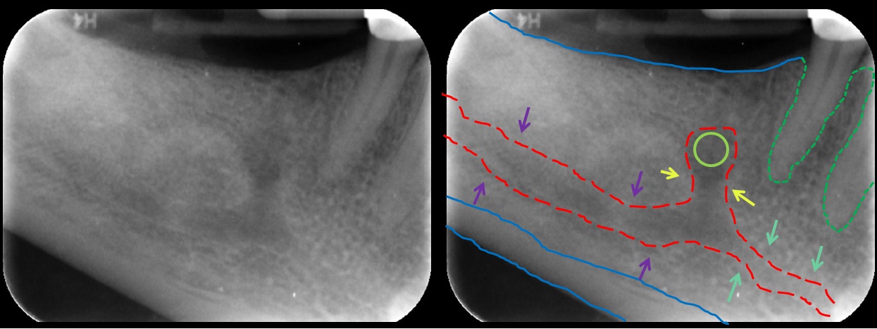
In human anatomy, the mandibular canal is a canal within the mandible that contains the inferior alveolar nerve, inferior alveolar artery, and inferior alveolar vein. It runs obliquely downward and forward in the ramus, and then horizontally forward in the body, where it is placed under the alveoli and communicates with them by small openings.
On arriving at the incisor teeth, it turns back to communicate with the mental foramen, giving off a small canal known as the mandibular incisive canal, which run to the cavities containing the incisor teeth. It carries branches of the inferior alveolar nerve and artery.
The mandibular canal is continuous with two foramina: the mental foramen which opens in the mental region of the mandible and carried the distal fibres of the inferior alveolar nerve as the mental nerve; and the mandibular foramen on medial aspect of ramus, into which the mandibular nerve enters to become the inferior alveolar nerve. The mandibular canal often runs close to the apices of the third molar tooth, and the inferior alveolar nerve can become damaged during removal of this tooth, causing sensory disturbance in the distribution of the nerve. This is sometimes the case for the second or first molar teeth, and care must be taken during removal or root canal treatment in such cases to prevent nerve injury or extrusion of root canal filling materials.
Variations
Several variations of the mandibular canal exist with varying frequency. The most common variant is the retromolar canal (~10 % of canals), whereby a branch is given off in the mandibular ramus which terminates in the retromolar region of the mandible. The retromolar canal may cause bleeding during surgery in the retromolar region such as removal of mandibular third molar teeth. Other variants include a bifid canal with a branch (~41%): following the course of the main mandibular canal before re-joining it (forward or buccolingual type); terminating at the apex of a tooth, usually the molar teeth (dental type); opening as an accessory mental foramen. A trifid mandibular canal variation has also been described.
Additional images
File:Slide2cec.JPG|Mandibular nerve and bone. Deep dissection. Anterior view. File:Slide7cece.JPG|Infratemporal fossa. Lingual and inferior alveolar nerve. Deep dissection. Anterolateral view
References
References
- Greenstein, G; Cavallaro, J; [[Dennis Tarnow. Tarnow, D]]: "Practical Application of Anatomy the Dental Implant Surgeon," ''J Perio'' October 2008, pg 1837
- "Yahoo | Mail, Weather, Search, Politics, News, Finance, Sports & Videos".
- (2023-08-14). "Prevalence and characteristics of accessory mandibular canals in an eastern Chinese population by cone beam computed tomography". Surgical and Radiologic Anatomy.
- (November 2017). "A prevalence study of bifid mandibular canals using cone beam computed tomography". Oral Surgery.
- (2014-09-01). "Assessment of bifid and trifid mandibular canals using cone-beam computed tomography". Imaging Science in Dentistry.
- (March 2012). "The clinical relevance of bifid and trifid mandibular canals". Oral and Maxillofacial Surgery.
This article was imported from Wikipedia and is available under the Creative Commons Attribution-ShareAlike 4.0 License. Content has been adapted to SurfDoc format. Original contributors can be found on the article history page.
Ask Mako anything about Mandibular canal — get instant answers, deeper analysis, and related topics.
Research with MakoFree with your Surf account
Create a free account to save articles, ask Mako questions, and organize your research.
Sign up freeThis content may have been generated or modified by AI. CloudSurf Software LLC is not responsible for the accuracy, completeness, or reliability of AI-generated content. Always verify important information from primary sources.
Report