From Surf Wiki (app.surf) — the open knowledge base
Mammary gland
Exocrine gland in humans and other mammals
Exocrine gland in humans and other mammals
| Field | Value |
|---|---|
| Name | Mammary gland |
| Image | Breast anatomy normal scheme.png |
| Caption | Cross-section of the human mammary gland: |
| Precursor | Mesoderm |
| (blood vessels and connective tissue) | |
| Ectoderm | |
| (cellular elements) | |
| Artery | Internal thoracic artery |
| Lateral thoracic artery | |
| Vein | Internal thoracic vein |
| Axillary vein | |
| Nerve | Supraclavicular nerves |
| Intercostal nerves | |
| (lateral and medial branches) | |
| Lymph | Pectoral axillary lymph nodes |
(blood vessels and connective tissue) Ectoderm (cellular elements) Lateral thoracic artery Axillary vein Intercostal nerves (lateral and medial branches)
A mammary gland is an exocrine gland that produces milk in mammals, including humans. Mammals get their name from the Latin word mamma, "breast". The mammary glands are arranged in organs such as the breasts in primates (for example, humans and chimpanzees), the udder in ruminants (for example, cows, goats, sheep, and deer), and the dugs of other animals (for example, dogs, cats, rabbits and pigs) to feed young offspring. Lactorrhea, the occasional production of milk by the glands, can occur in any mammal, but in most mammals, lactation, the production of enough milk for nursing, occurs only in phenotypic females who have gestated in recent months or years. It is directed by hormonal guidance from sex steroids. In a few mammalian species, male lactation can occur. With humans, male lactation can occur only under specific circumstances.
Mammals are divided into 3 groups: monotremes, metatherians, and eutherians. In the case of monotremes, their mammary glands are modified sebaceous glands and without nipples. Concerning most metatherians and eutherians, only females have functional mammary glands, with the exception of some bat species. Their mammary glands can be termed as breasts or udders. In the case of breasts, each mammary gland has its own nipple (e.g., human mammary glands). In the case of udders, pairs of mammary glands comprise a single mass, with more than one nipple (or teat) hanging from it. For instance, cows and buffalo udders have two pairs of mammary glands and four teats, whereas sheep and goat udders have one pair of mammary glands with two teats protruding from the udder. Each mammary gland produces milk for a single teat and is evolutionarily derived from modified sweat glands.
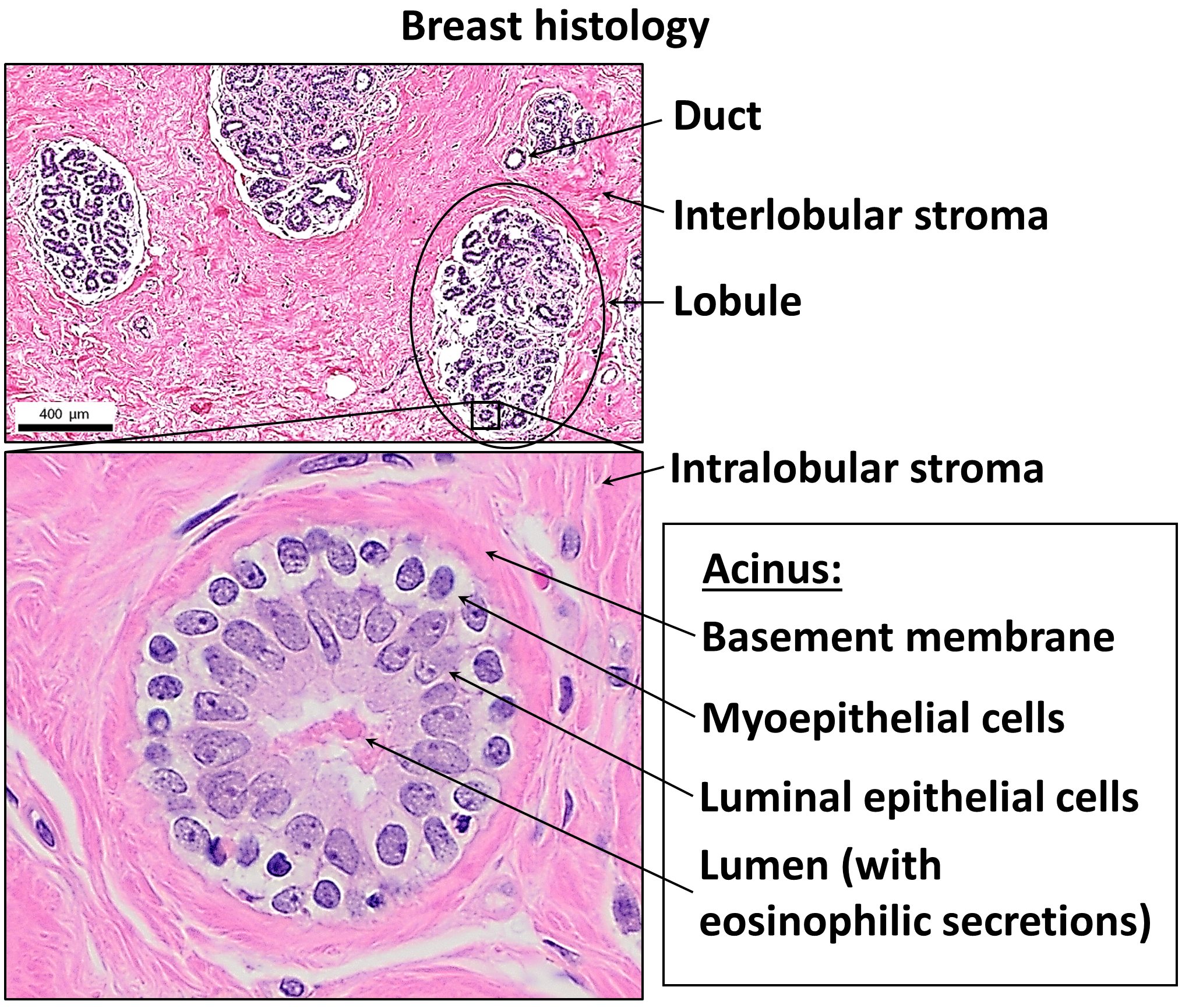
Structure
The basic components of a mature mammary gland are the alveoli (hollow cavities, a few millimeters large), which are lined with milk-secreting cuboidal cells and surrounded by myoepithelial cells. These alveoli join to form groups known as lobules. Each lobule has a lactiferous duct that drains into openings in the nipple. The myoepithelial cells contract under the stimulation of oxytocin, excreting the milk secreted by alveolar units into the lobule lumen toward the nipple. As the infant begins to suck, the oxytocin-mediated "let down reflex" ensues, and the mother's milk is secreted—not sucked—from the gland into the infant's mouth.{{cite journal
All the milk-secreting tissue leading to a single lactiferous duct is collectively called a "simple mammary gland"; in a "complex mammary gland", all the simple mammary glands serve one nipple. Humans normally have two complex mammary glands, one in each breast, and each complex mammary gland consists of 10–20 simple glands. The opening of each simple gland on the surface of the nipple is called a "pore." The presence of more than two nipples is known as polythelia and the presence of more than two complex mammary glands as polymastia.
Maintaining the correct polarized morphology of the lactiferous duct tree requires another essential component – mammary epithelial cells extracellular matrix (ECM) which, together with adipocytes, fibroblast, inflammatory cells, and others, constitute mammary stroma.{{Cite journal | doi-access =
Histology
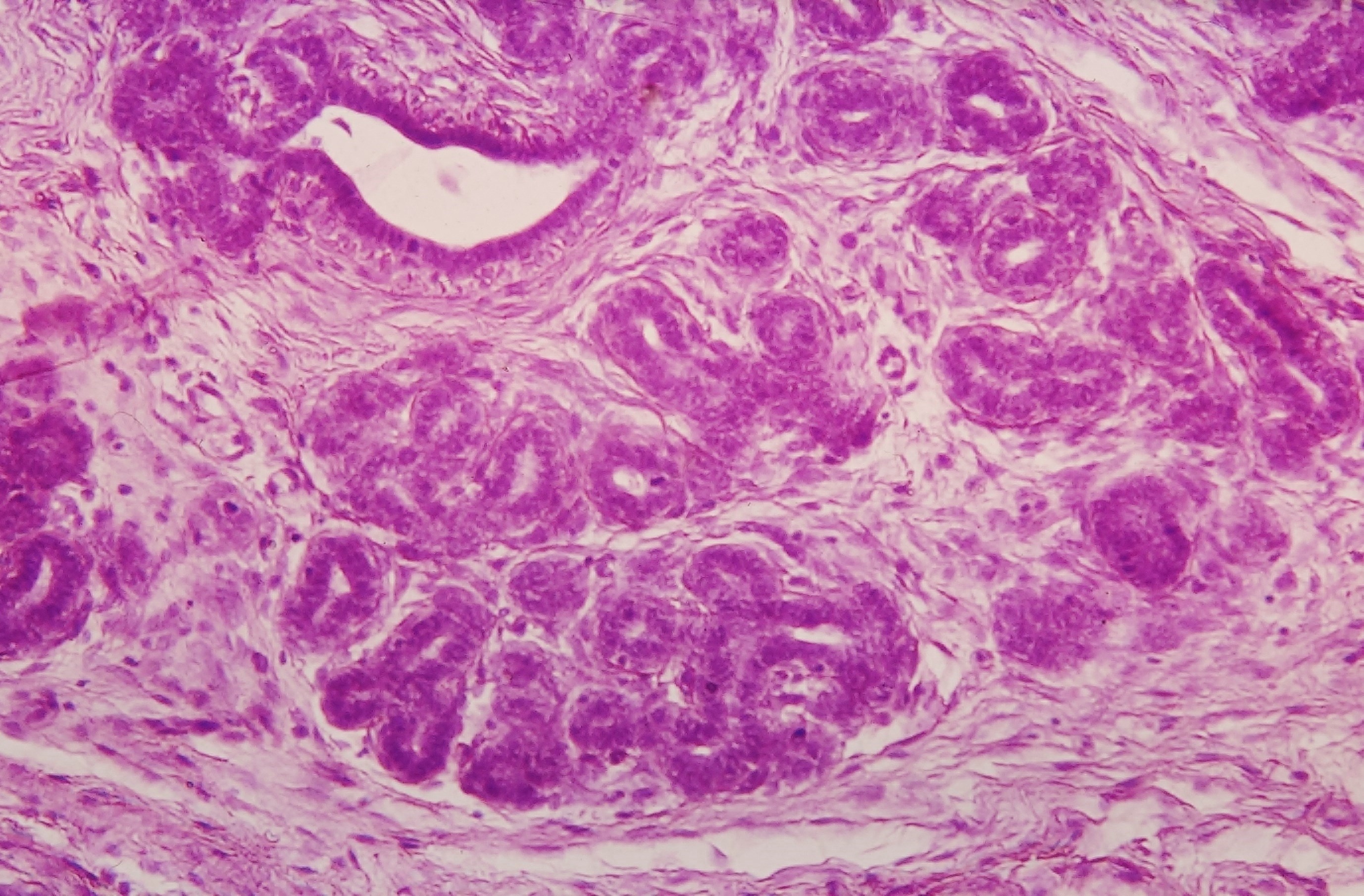
A mammary gland is a specific type of apocrine gland specialized for manufacture of colostrum (first milk) when giving birth. Mammary glands can be identified as apocrine because they exhibit striking "decapitation" secretion. Many sources assert that mammary glands are modified sweat glands.
Development
Mammary glands develop during different growth cycles. They exist in both sexes during the embryonic stage, forming only a rudimentary duct tree at birth. In this stage, mammary gland development depends on systemic (and maternal) hormones, but is also under the (local) regulation of paracrine communication between neighboring epithelial and mesenchymal cells by parathyroid hormone-related protein (PTHrP).{{Cite journal | doi-access = free
Biochemistry
Estrogen and growth hormone (GH) are essential for the ductal component of mammary gland development, and act synergistically to mediate it. Neither estrogen nor GH are capable of inducing ductal development without the other. The role of GH in ductal development has been found to be mostly mediated by its induction of the secretion of insulin-like growth factor 1 (IGF-1), which occurs both systemically (mainly originating from the liver) and locally in the mammary fat pad through activation of the growth hormone receptor (GHR). However, GH itself also acts independently of IGF-1 to stimulate ductal development by upregulating estrogen receptor (ER) expression in mammary gland tissue, which is a downstream effect of mammary gland GHR activation. In any case, unlike IGF-1, GH itself is not essential for mammary gland development, and IGF-1 in conjunction with estrogen can induce normal mammary gland development without the presence of GH. In addition to IGF-1, other paracrine growth factors such as epidermal growth factor (EGF), transforming growth factor beta (TGF-β), amphiregulin, fibroblast growth factor (FGF), and hepatocyte growth factor (HGF) are involved in breast development as mediators downstream to sex hormones and GH/IGF-1.
During embryonic development, IGF-1 levels are low, and gradually increase from birth to puberty. At puberty, the levels of GH and IGF-1 reach their highest levels in life and estrogen begins to be secreted in high amounts in females, which is when ductal development mostly takes place. Under the influence of estrogen, stromal and fat tissue surrounding the ductal system in the mammary glands also grows. After puberty, GH and IGF-1 levels progressively decrease, which limits further development until pregnancy, if it occurs. During pregnancy, progesterone and prolactin are essential for mediating lobuloalveolar development in estrogen-primed mammary gland tissue, which occurs in preparation of lactation and nursing.
Androgens such as testosterone inhibit estrogen-mediated mammary gland development (e.g., by reducing local ER expression) through activation of androgen receptors expressed in mammary gland tissue, and in conjunction with relatively low estrogen levels, are the cause of the lack of developed mammary glands in males.
Timeline
Before birth
Mammary gland development is characterized by the unique process by which the epithelium invades the stroma. The development of the mammary gland occurs mainly after birth. During puberty, tubule formation is coupled with branching morphogenesis which establishes the basic arboreal network of ducts emanating from the nipple.
Developmentally, mammary gland epithelium is constantly produced and maintained by rare epithelial cells, dubbed as mammary progenitors which are ultimately thought to be derived from tissue-resident stem cells.
Embryonic mammary gland development can be divided into a series of specific stages. Initially, the formation of the milk lines that run between the fore and hind limbs bilaterally on each side of the midline occurs around embryonic day 10.5 (E10.5). The second stage occurs at E11.5 when placode formation begins along the mammary milk line. This will eventually give rise to the nipple. Lastly, the third stage occurs at E12.5 and involves the invagination of cells within the placode into the mesenchyme, leading to a mammary anlage (biology).
The primitive (stem) cells are detected in embryo and their numbers increase steadily during development
Growth
Postnatally, the mammary ducts elongate into the mammary fat pad. Then, starting around four weeks of age, mammary ductal growth increases significantly with the ducts invading towards the lymph node. Terminal end buds, the highly proliferative structures found at the tips of the invading ducts, expand and increase greatly during this stage. This developmental period is characterized by the emergence of the terminal end buds and lasts until an age of about 7–8 weeks.
By the pubertal stage, the mammary ducts have invaded to the end of the mammary fat pad. At this point, the terminal end buds become less proliferative and decrease in size. Side branches form from the primary ducts and begin to fill the mammary fat pad. Ductal development decreases with the arrival of sexual maturity and undergoes estrous cycles (proestrus, estrus, metestrus, and diestrus). As a result of estrous cycling, the mammary gland undergoes dynamic changes where cells proliferate and then regress in an ordered fashion.
Pregnancy
During pregnancy, the ductal systems undergo rapid proliferation and form alveolar structures within the branches to be used for milk production. After delivery, lactation occurs within the mammary gland; lactation involves the secretion of milk by the luminal cells in the alveoli. Contraction of the myoepithelial cells surrounding the alveoli will cause the milk to be ejected through the ducts and into the nipple for the nursing infant. Upon weaning of the infant, lactation stops and the mammary gland turns in on itself, a process called involution. This process involves the controlled collapse of mammary epithelial cells where cells begin apoptosis in a controlled manner, reverting the mammary gland back to a pubertal state.
Postmenopausal
During postmenopause, due to much lower levels of estrogen, and due to lower levels of GH and IGF-1, which decrease with age, mammary gland tissue atrophies and the mammary glands become smaller.
Physiology
Hormonal control
Lactiferous duct development occurs in females in response to circulating hormones. First development is frequently seen during pre- and postnatal stages, and later during puberty. Estrogen promotes branching differentiation,{{Cite journal | article-number = 201 | doi-access = free | doi-access = free
There is preliminary evidence that soybean intake mildly stimulates the breast glands in pre- and postmenopausal women.
Pregnancy
Secretory alveoli develop mainly in pregnancy, when rising levels of prolactin, estrogen, and progesterone cause further branching, together with an increase in adipose tissue and a richer blood flow. In gestation, serum progesterone remains at a stably high concentration so signaling through its receptor is continuously activated. As one of the transcribed genes, Wnts secreted from mammary epithelial cells act paracrinely to induce more neighboring cells' branching.{{Cite journal | doi-access = free
Weaning
During weaning, decreased prolactin, missing mechanical stimulation (baby suckling), and changes in osmotic balance caused by milk stasis and leaking of tight junctions cause cessation of milk production. It is the (passive) process of a child or animal ceasing to be dependent on the mother for nourishment. In some species there is complete or partial involution of alveolar structures after weaning, in humans there is only partial involution and the level of involution in humans appears to be highly individual. The glands in the breast do secrete fluid also in nonlactating women. In some other species (such as cows), all alveoli and secretory duct structures collapse by programmed cell death (apoptosis) and autophagy for lack of growth promoting factors either from the ECM or circulating hormones.{{Cite journal
Clinical significance
Tumorigenesis in mammary glands can be induced biochemically by abnormal expression level of circulating hormones or local ECM components,{{Cite journal | article-number = 11 | doi-access = free
Other mammals
General
The breasts of female humans vary from most other mammals that tend to have less conspicuous mammary glands. The number and positioning of mammary glands varies widely in different mammals. The protruding teats and accompanying glands can be located anywhere along the two milk lines. In general most mammals develop mammary glands in pairs along these lines, with a number approximating the number of young typically birthed at a time. The number of teats varies from 2 (in most primates) to 18 (in pigs). The Virginia opossum has 13, one of the few mammals with an odd number. The following table lists the number and position of teats and glands found in a range of mammals:
| Species | Anterior | |||
|---|---|---|---|---|
| (thoracic) | Intermediate | |||
| (abdominal) | Posterior | |||
| (inguinal) | Total | |||
| Goat, sheep, horse | ||||
| guinea pig | 0 | 0 | 2 | 2 |
| Cattle | 0 | 0 | 4 | 4 |
| Cat | 2 | 2 | 4 | 8 |
| Dog | 4 | 2 | 2 or 4 | 8 or 10 |
| Mouse | 6 | 0 | 4 | 10 |
| Rat | 6 | 2 | 4 | 12 |
| Pig | 6 | 6 | 6 | 18 |
| Proboscideans, primates | 2 | 0 | 0 | 2 |
| Virginia opossum | 0 | 0 | 13 | 13 |
| Southern red-sided opossum | 0 | 0 | 25 to 27 | 25 to 27 |
Male mammals typically have rudimentary mammary glands and nipples, with a few exceptions: male mice do not have nipples, male marsupials do not have mammary glands, and male horses lack nipples. The male dayak fruit bat has lactating mammary glands. Male lactation occurs infrequently in some species.
Mammary glands are true protein factories, and several labs have constructed transgenic animals, mainly goats and cows, to produce proteins for pharmaceutical use. Complex glycoproteins such as monoclonal antibodies or antithrombin cannot be produced by genetically engineered bacteria, and the production in live mammals is much cheaper than the use of mammalian cell cultures.
Evolution
There are many theories on how mammary glands evolved. For example, it is thought that the mammary gland is a transformed sweat gland, more closely related to apocrine sweat glands.{{Cite journal
Lactation is thought to have developed long before the evolution of the mammary gland and mammals; see evolution of lactation.
Additional images
Image:illu_breast_anatomy.jpg|Cross section of the breast of a human female Image:Säugende Hündin.JPG|Dog Image:Bezerro mamando REFON.jpg|Cattle Image:White Cat Nursing Four Kittens HQ.jpg|Cat Image:Piglets1.jpg|Pig Image:Goat kid feeding on mothers milk.jpg|Goat Image:Elephant_breastfeading.jpg|Elephant Camelus dromedarius Euter Zoo Landau Juni 2011.JPG|Dromedary camel Netzgiraffe Euter.JPG|Giraffe
References
Bibliography
- Moore, Keith L. et al. (2010) Clinically Oriented Anatomy 6th Ed
References
- Gray, Henry. (1918). "Anatomy of the Human Body".
- (2010-09-30). "Breastfeeding: A Guide for the Medical Profession". Mosby/Elsevier.
- (1 December 2006). "Anatomy of the Thoracic Wall, Axilla and Breast". International Journal of Morphology.
- (February 2016). "Anatomy of the nipple and breast ducts". Gland Surgery.
- (2018). "Clinically oriented anatomy". Wolters Kluwer.
- Ackerman (2005) ch.1 [http://www.derm101.com/content/13501 ''Apocrine Units''] {{webarchive. link. (21 April 2011)
- Krstic, Radivoj V.. (18 March 2004). "Human Microscopic Anatomy: An Atlas for Students of Medicine and Biology". Springer.
- (2016-10-01). "Modeling mammary organogenesis from biological first principles: Cells and their physical constraints". Progress in Biophysics and Molecular Biology.
- (2 December 2010). "Hormone Action in the Mammary Gland". Cold Spring Harbor Perspectives in Biology.
- (1998). "Role of IGF-I in normal mammary development". Breast Cancer Res. Treat..
- (1997). "Early mammary development: growth hormone and IGF-1". J Mammary Gland Biol Neoplasia.
- (1999). "Insulin-like growth factor I is essential for terminal end bud formation and ductal morphogenesis during mammary development". Endocrinology.
- (2000). "IGF-I: an essential factor in terminal end bud formation and ductal morphogenesis". J Mammary Gland Biol Neoplasia.
- (2008). "IGF-I, GH, and sex steroid effects in normal mammary gland development". J Mammary Gland Biol Neoplasia.
- (2005). "Mouse models of transforming growth factor beta impact in breast development and cancer". Endocr. Relat. Cancer.
- (2007). "Estrogen regulation of mammary gland development and breast cancer: amphiregulin takes center stage". Breast Cancer Res..
- (2011). "Hepatocyte growth factor profile with breast cancer". Indian J Pathol Microbiol.
- (2011). "Anatomy and Physiology for Midwives". Elsevier Health Sciences.
- (2010). "Mammary Gland Growth Factors: Roles in Normal Development and in Cancer". Cold Spring Harbor Perspectives in Biology.
- (28 March 2012). "Diseases of the Breast". Lippincott Williams & Wilkins.
- (2007). "The potential clinical applications of insulin-like growth factor-1 ligand in human breast cancer". Anticancer Res..
- Leonard R. Johnson. (2003). "Essential Medical Physiology". Academic Press.
- (1997). "Breast size in relation to endogenous hormone levels, body constitution, and oral contraceptive use in healthy nulligravid women aged 19–25 years". Am. J. Epidemiol..
- (2000). "Testosterone inhibits estrogen-induced mammary epithelial proliferation and suppresses estrogen receptor expression". FASEB J..
- (2013). "Gynecomastia in adolescent males". Semin Plast Surg.
- (Sep 1967). "Studies of mouse mammary glands. I. Cytomorphology of the normal mammary gland". J Natl Cancer Inst.
- (2019-07-15). "Mammary stem cells and progenitors: targeting the roots of breast cancer for prevention". The EMBO Journal.
- Hens, JR. (10 August 2005). "Key stages of mammary gland development: molecular mechanisms involved in the formation of the embryonic mammary gland". Breast Cancer Res..
- Makarem, M. (Apr 2013). "Stem Cells and the Developing Mammary Gland". J Mammary Gland Biol Neoplasia.
- Daniel, CW. (January 1999). "The mammary gland: a model for development". Journal of Mammary Gland Biology and Neoplasia.
- Kurzer MS. (March 2002). "Hormonal effects of soy in premenopausal women and men". The Journal of Nutrition.
- (1975). "Association of Race, Age, Menopausal Status, and Cerumen Type With Breast Fluid Secretion in Nonlactating Women, as Determined by Nipple Aspiration". Journal of the National Cancer Institute.
- "With the Wild Things – Transcripts". Digitalcollections.fiu.edu.
- Stockard, Mary (2005) [https://web.archive.org/web/20100701012225/http://www.awrc.org/Baby%20Opossums.htm Raising Orphaned Baby Opossums]. Alabama Wildlife Center.
- (2005). "Animal Science and Industry". Pearson Prentice Hall.
- Dog breeds vary in the number of mammary glands: larger breeds tend to have 5 pairs, smaller breeds have 4 pairs.{{citation needed. (December 2017)
- P Smith 2008 [http://www.faunaparaguay.com/mamm26Monodelphissorex.pdf Red-Sided Short-Tailed Opossum]. Fauna Paraguay
- (November 2008). "Conversion of the Nipple to Hair-Bearing Epithelia by Lowering Bone Morphogenetic Protein Pathway Activity at the Dermal-Epidermal Interface". Am J Pathol.
- (17 August 2006). "Marsupials". Cambridge University Press.
- (2021). "Development and Pathology of the Equine Mammary Gland". Journal of Mammary Gland Biology and Neoplasia.
- (1994). "Lactation in male fruit bats". Nature.
- (2009). "Male lactation: why, why not and is it care?". Trends in Ecology & Evolution.
- (2009-08-01). "Lactating Porcine Mammary Tissue Catabolizes Branched-Chain Amino Acids for Glutamine and Aspartate Synthesis". The Journal of Nutrition.
- (17 January 2012). "BBC News – The goats with spider genes and silk in their milk". bbc.co.uk.
- [https://web.archive.org/web/20090419024229/http://nationalzoo.si.edu/ConservationAndScience/SpotlightOnScience/oftedalolav20030714.cfm Lactating on Eggs]. Smithsonian National Zoo, 14 July 2003.
- [https://web.archive.org/web/20070312005054/http://scienceblogs.com/pharyngula/2006/05/breast_beginnings.php Breast beginnings]. scienceblogs.com
- (1983). "A Role for Aggregation Pheromones in the Evolution of Mammallike Reptile Lactation". The American Naturalist.
This article was imported from Wikipedia and is available under the Creative Commons Attribution-ShareAlike 4.0 License. Content has been adapted to SurfDoc format. Original contributors can be found on the article history page.
Ask Mako anything about Mammary gland — get instant answers, deeper analysis, and related topics.
Research with MakoFree with your Surf account
Create a free account to save articles, ask Mako questions, and organize your research.
Sign up freeThis content may have been generated or modified by AI. CloudSurf Software LLC is not responsible for the accuracy, completeness, or reliability of AI-generated content. Always verify important information from primary sources.
Report