From Surf Wiki (app.surf) — the open knowledge base
Left ventricular hypertrophy

| Field | Value |
|---|---|
| name | Left ventricular hypertrophy |
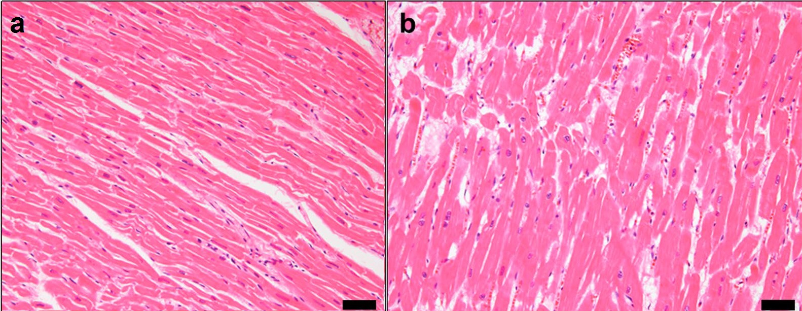
| image | Heart left ventricular hypertrophy sa.jpg |
| caption | A heart with left ventricular hypertrophy in short-axis view |
| field | Cardiology |
| complications | Hypertrophic cardiomyopathy, Heart failure |
| diagnosis | Echocardiography, cardiovascular MRI |
| differential | Athletic heart syndrome |
Left ventricular hypertrophy (LVH) is thickening of the heart muscle of the left ventricle of the heart, that is, left-sided ventricular hypertrophy and resulting increased left ventricular mass.
Causes
While ventricular hypertrophy occurs naturally as a reaction to aerobic exercise and strength training, it is most frequently referred to as a pathological reaction to cardiovascular disease, or high blood pressure. It is one aspect of ventricular remodeling.
While LVH itself is not a disease, it is usually a marker for disease involving the heart. Disease processes that can cause LVH include any disease that increases the afterload that the heart has to contract against, and some primary diseases of the muscle of the heart. Causes of increased afterload that can cause LVH include aortic stenosis, aortic insufficiency and hypertension. Primary disease of the muscle of the heart that cause LVH are known as hypertrophic cardiomyopathies, which can lead into heart failure.
Long-standing mitral insufficiency also leads to LVH as a compensatory mechanism.
LV mass increases with ageing.
Associated genes include OGN (osteoglycin).
Diagnosis
The commonly used method to diagnose LVH is echocardiography, with which the thickness of the muscle of the heart can be measured. The electrocardiogram (ECG) often shows signs of increased voltage from the heart in individuals with LVH, so this is often used as a screening test to determine who should undergo further testing.
Echocardiography
| Severe | 17 mm |
|---|
Two dimensional echocardiography can produce images of the left ventricle. The thickness of the left ventricle as visualized on echocardiography correlates with its actual mass. Left ventricular mass can be further estimated based on geometric assumptions of ventricular shape using the measured wall thickness and internal diameter. Average thickness of the left ventricle, with numbers given as 95% prediction interval for the short axis images at the mid-cavity level are:
- Women: 4 – 8 mm
- Men: 5 – 9 mm
CT & MRI
CT and MRI-based measurement can be used to measure the left ventricle in three dimensions and calculate left ventricular mass directly. MRI based measurement is considered the "gold standard" for left ventricular mass, though is usually not readily available for common practice. In older individuals, age related remodeling of the left ventricle's geometry can lead to a discordancy between CT and echocardiographic based measurements of left ventricular mass.
ECG criteria

There are several sets of criteria used to diagnose LVH via electrocardiography. None of them are perfect, though by using multiple criteria sets, the sensitivity and specificity are increased.
The Sokolow-Lyon index:
- S in V1 + R in V5 or V6 (whichever is larger) ≥ 35 mm (≥ 7 large squares)
- R in aVL ≥ 11 mm
The Cornell voltage criteria for the ECG diagnosis of LVH involve measurement of the sum of the R wave in lead aVL and the S wave in lead V3. The Cornell criteria for LVH are:
- S in V3 + R in aVL 28 mm (men)
- S in V3 + R in aVL 20 mm (women)
The Romhilt-Estes point score system ("diagnostic" 5 points; "probable" 4 points):
| Delayed intrinsicoid deflection in V5 or V6 (0.05 sec) | 1 |
|---|
Other voltage-based criteria for LVH include:
- Lead I: R wave 14 mm
- Lead aVR: S wave 15 mm
- Lead aVL: R wave 12 mm
- Lead aVF: R wave 21 mm
- Lead V5: R wave 26 mm
- Lead V6: R wave 20 mm
Diagnostic accuracy of electrocardiography in left ventricular hypertrophy can be enhanced with artificial intelligence analysis.
Treatment
Treatment is typically focused on resolving the cause of the LVH with the enlargement not permanent in all cases. In some cases the growth can regress with the reduction of blood pressure.
LVH may be a factor in determining treatment or diagnosis for other conditions, for example, LVH is used in the staging and risk stratification of Non-ischemic cardiomyopathies such as Fabry's Disease. Patients with LVH may have to participate in more complicated and precise diagnostic procedures, such as echocardiography or cardiac MRI.
References
References
- (January 2013). "Hypertrophic cardiomyopathy". Elsevier BV.
- "Ask the doctor: Left Ventricular Hypertrophy".
- (2007). "Rationale and design of the SMART Heart study: A prediction model for left ventricular hypertrophy in hypertension". Netherlands Heart Journal.
- (May 2008). "Integrated genomic approaches implicate osteoglycin (Ogn) in the regulation of left ventricular mass". Nature Genetics.
- (March 2008). "Use of cardiac allografts with mild and moderate left ventricular hypertrophy can be safely used in heart transplantation to expand the donor pool". Journal of the American College of Cardiology.
- (January 2015). "Recommendations for cardiac chamber quantification by echocardiography in adults: an update from the American Society of Echocardiography and the European Association of Cardiovascular Imaging". Journal of the American Society of Echocardiography.
- (July 2012). "Normal left ventricular myocardial thickness for middle-aged and older subjects with steady-state free precession cardiac magnetic resonance: the multi-ethnic study of atherosclerosis". Circulation: Cardiovascular Imaging.
- (March 2002). "Assessment of left ventricular mass by cardiovascular magnetic resonance". Hypertension.
- (2019-10-24). "Echocardiography overestimates LV mass in the elderly as compared to cardiac CT". PLOS ONE.
- "Lesson VIII - Ventricular Hypertrophy".
- (February 1949). "The ventricular complex in left ventricular hypertrophy as obtained by unipolar precordial and limb leads". American Heart Journal.
- (April 1998). "Time-voltage QRS area of the 12-lead electrocardiogram: detection of left ventricular hypertrophy". Hypertension.
- (March 1987). "Improved sex-specific criteria of left ventricular hypertrophy for clinical and computer interpretation of electrocardiograms: validation with autopsy findings". Circulation.
- (2023). "Current and Future Use of Artificial Intelligence in Electrocardiography". Journal of Cardiovascular Development and Disease.
- (2006). "From left ventricular hypertrophy to congestive heart failure: management of hypertensive heart disease". Progress in Cardiovascular Diseases.
- (November 2019). "Multimodality Imaging Assessment of Fabry Disease". Circulation: Cardiovascular Imaging.
- "Five Things Physicians and Patients Should Question". American Society of Nuclear Cardiology.
- (August 2007). "ACC/AHA 2007 guidelines for the management of patients with unstable angina/non-ST-Elevation myocardial infarction: a report of the American College of Cardiology/American Heart Association Task Force on Practice Guidelines (Writing Committee to Revise the 2002 Guidelines for the Management of Patients With Unstable Angina/Non-ST-Elevation Myocardial Infarction) developed in collaboration with the American College of Emergency Physicians, the Society for Cardiovascular Angiography and Interventions, and the Society of Thoracic Surgeons endorsed by the American Association of Cardiovascular and Pulmonary Rehabilitation and the Society for Academic Emergency Medicine". Journal of the American College of Cardiology.
This article was imported from Wikipedia and is available under the Creative Commons Attribution-ShareAlike 4.0 License. Content has been adapted to SurfDoc format. Original contributors can be found on the article history page.
Ask Mako anything about Left ventricular hypertrophy — get instant answers, deeper analysis, and related topics.
Research with MakoFree with your Surf account
Create a free account to save articles, ask Mako questions, and organize your research.
Sign up freeThis content may have been generated or modified by AI. CloudSurf Software LLC is not responsible for the accuracy, completeness, or reliability of AI-generated content. Always verify important information from primary sources.
Report