From Surf Wiki (app.surf) — the open knowledge base
Intraductal papillary mucinous neoplasm

| Field | Value |
|---|---|
| name | Intraductal papillary mucinous neoplasm |
| synonyms | IPMN |
| image | IPMN T2w ax-07 a.jpg |
| caption | Intraductal papillary mucinous neoplasm in magnetic resonance imaging. |
| field | Gastroenterology |
| symptoms | Usually asymptomatic |
| onset | 50-70 years of age |
| types | Main duct, branch duct or mixed |
| risks | Male gender |
| differential | Mucinous cystic neoplasm |
| treatment | Imaging surveillance, |
| surgical resection |
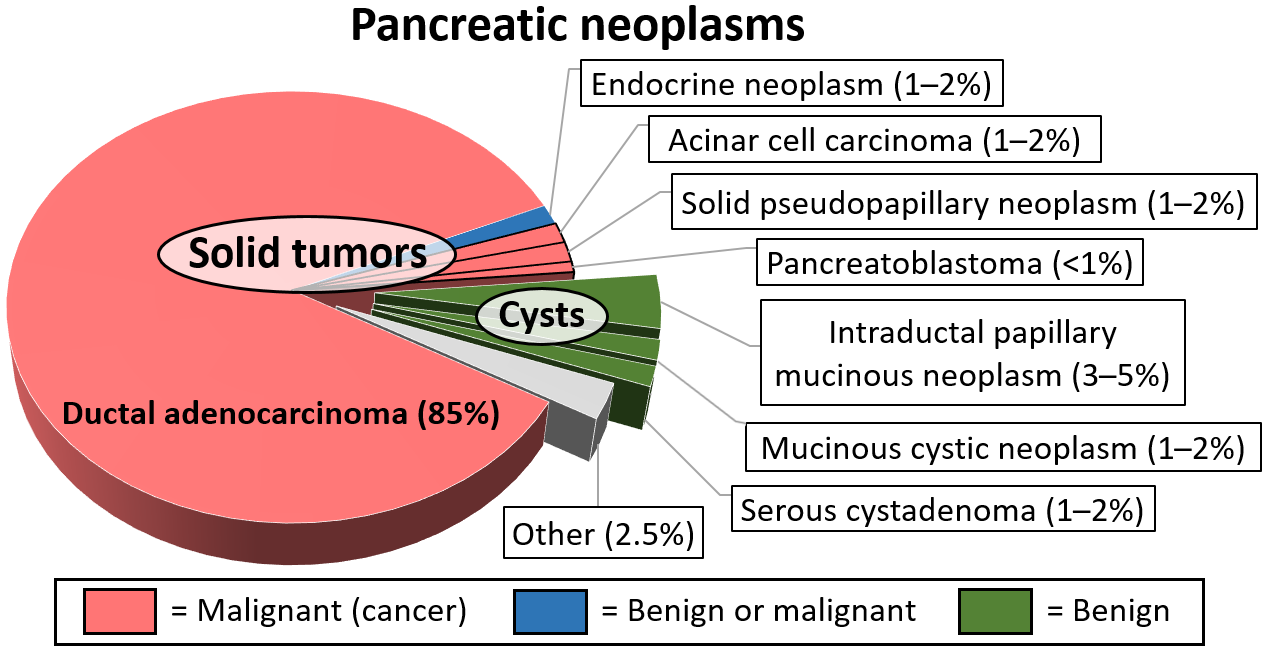
surgical resection Intraductal papillary mucinous neoplasm (IPMN) is a type of tumor that can occur within the cells of the pancreatic duct. IPMN tumors produce mucus, and this mucus can form pancreatic cysts. Although intraductal papillary mucinous neoplasms are benign tumors, they can progress to pancreatic cancer. As such IPMN is viewed as a precancerous condition. Once an intraductal papillary mucinous neoplasm has been found, the management options include close monitoring and pre-emptive surgery.
Histology
IPMNs are lined with mucin-secreting columnar epithelium.
Diagnosis
In most cases, IPMNs are diagnosed based on clinical and radiographic criteria. If fluid from the cyst is aspirated, the CEA level is typically elevated. Confirmation of the diagnosis with tissue is rarely necessary.
By histopathology, IPMN is characterized on light microscopy by Mucinous epithelial cells, and growth within the pancreatic ducts. Mucin 5AC is a useful immunohistochemistry marker. Characteristic genetic alterations are those of KRAS and GNAS.
Further subtyping of IPMN can be done as either:
- Gross pathology: Main duct, branch duct, and mixed duct lesions, which determines surgical management. Main duct lesion is the segmental or diffuse dilatation of main pancreatic duct greater than 5 mm without other causes of obstruction. Meanwhile, branch duct lesion is the pancreatic cyst more than 5 mm that communicates with the main duct. The mixed duct lesions fulfills both criteria above.
- By light microscopy and immunohistochemistry: Gastric, intestinal, pancreaticbiliary, and oncocytic.
Treatment
The treatment of choice for main-duct IPMNs is resection due to approximately 50% chance of malignancy. Side-branch IPMNs are occasionally monitored with regular CT or MRIs, but most are eventually resected, with a 30% rate of malignancy in these resected tumors. Indications for surgical resection include obstructive jaundice, an enhancing mural nodule 5 mm, and pancreatic duct dilation (10 mm).
Surgery can include the removal of the head of the pancreas (a pancreaticoduodenectomy), removal of the body and tail of the pancreas (a distal pancreatectomy), or rarely removal of the entire pancreas (a total pancreatectomy). In selected cases the surgery can be performed using minimally invasive techniques such as laparoscopy or robotic surgery. A study using Surveillance, Epidemiology, and End Results Registry (SEER) data suggested that increased lymph node counts harvested during the surgery were associated with better survival in invasive IPMN patients.
Prognosis
Survival 5 years after resection of an IPMN without malignancy is approximately 80%, 85% with malignancy but no lymph node spread and 0% with malignancy spreading to lymph nodes.
Epidemiology
Side branch IPMNs are the most common pancreatic cysts. IPMNs occur more often in men than women, and often occur in the 6th and 7th decade of life.
History
In 1982, IPMN was reported as a "mucin-producing tumor" by Kazuhiko Ohashi of the Japanese Foundation for Cancer Research.
References
References
- "Intraductal Papillary Mucinous Neoplasms of the Pancreas". [[Johns Hopkins University]].
- (March 2015). "Imaging patterns of intraductal papillary mucinous neoplasms of the pancreas: an illustrated discussion of the International Consensus Guidelines for the Management of IPMN". Abdominal Imaging.
- "Precancerous conditions of the pancreas - Canadian Cancer Society".
- (2017). "A resected case of two branch duct-type intraductal papillary mucinous neoplasms showing different clinical courses after a two-year follow-up". Clinical Journal of Gastroenterology.
- Diana Agostini-Vulaj. "Pancreas - Exocrine tumors / carcinomas - Intraductal papillary mucinous neoplasm (IPMN)".
- (2016). "Pathologic Evaluation and Reporting of Intraductal Papillary Mucinous Neoplasms of the Pancreas and Other Tumoral Intraepithelial Neoplasms of Pancreatobiliary Tract". Annals of Surgery.
- Pishvaian MJ, Brody JR. (2017). "Therapeutic Implications of Molecular Subtyping for Pancreatic Cancer.". Oncology (Williston Park).
- (2010). "Differences between main-duct and branch-duct intraductal papillary mucinous neoplasms of the pancreas". World Journal of Gastrointestinal Surgery.
- (2017-08-03). "Intraductal papillary mucinous neoplasm of the pancreas – epidemiology, risk factors, diagnosis, and management". Scandinavian Journal of Gastroenterology.
- (2007). "Characteristic Clinicopathological Features of the Types of Intraductal Papillary-Mucinous Neoplasms of the Pancreas". Pancreas.
- (October 2017). "Revisions of international consensus Fukuoka guidelines for the management of IPMN of the pancreas.". Pancreatology.
- "Pancreatic Surgery". [[Johns Hopkins University]].
- "Laproscopic Pancreatic Surgery". [[Johns Hopkins University]].
- Wu, WM. (Sep 2014). "An increased total resected lymph node count benefits survival following pancreas invasive intraductal papillary mucinous neoplasms resection: an analysis using the surveillance, epidemiology, and end result registry database.". PLOS ONE.
- Sohn, Taylor. (June 2004). "Intraductal Papillary Mucinous Neoplasms of the Pancreas". Annals of Surgery.
- Wang Y, Miller FH, Chen ZE, Merrick L, Mortele KJ, Hoff FL. (2011). "Diffusion-weighted MR imaging of solid and cystic lesions of the pancreas.". Radiographics.
- (April 2018). "ACG Clinical Guideline: Diagnosis and Management of Pancreatic Cysts.". The American Journal of Gastroenterology.
This article was imported from Wikipedia and is available under the Creative Commons Attribution-ShareAlike 4.0 License. Content has been adapted to SurfDoc format. Original contributors can be found on the article history page.
Ask Mako anything about Intraductal papillary mucinous neoplasm — get instant answers, deeper analysis, and related topics.
Research with MakoFree with your Surf account
Create a free account to save articles, ask Mako questions, and organize your research.
Sign up freeThis content may have been generated or modified by AI. CloudSurf Software LLC is not responsible for the accuracy, completeness, or reliability of AI-generated content. Always verify important information from primary sources.
Report