From Surf Wiki (app.surf) — the open knowledge base
Histiocytoma (dog)
Benign tumor in dogs

Benign tumor in dogs

A histiocytoma in the dog is a benign tumor. It is an abnormal growth in the skin of histiocytes (histiocytosis), a cell that is part of the immune system. A similar disease in humans, Hashimoto-Pritzker disease, is also a Langerhans cell histiocytosis. Dog breeds that may be more at risk for this tumor include Bulldogs, American Pit Bull Terriers, American Staffordshire Terriers, Scottish Terriers, Greyhounds, Boxers, and Boston Terriers. They also rarely occur in goats and cattle.
Histiocytic disorders
A histiocyte is a differentiated tissue cell that has its origin in the bone marrow. The source for histiocytes is the monocyte/macrophage line. Monocytes (found in the blood) and macrophages (found in tissue) are responsible for phagocytosis (ingestion) of foreign material in the body. Langerhans cells are dendritic cells found in the skin and function by internalizing antigens (foreign particles) and presenting them to T cells. They arise from monocytes. Histiocytic disorders refer to diseases that are caused by abnormal behavior of these cells. They include the following:
- Reactive diseases of Langerhans cells
- Histiocytomas
- Cutaneous histiocytosis
- Systemic histiocytosis
- Reactive diseases of macrophages
- Hemophagocytic syndrome - a condition where macrophages phagocytose myeloid and erythroid precursors (similar to hemophagocytic lymphohistiocytosis in humans)
- Malignant diseases of Langerhans cells
- Malignant histiocytosis - a condition found in Bernese Mountain Dogs
- Diffuse histiocytic sarcoma
- Localized histiocytic sarcoma
- Malignant diseases of macrophages
- Histiocytic lymphoma
Tumor biology
A histiocytoma originates from epidermal Langerhans cells of antigen-presenting cell lineage. Spontaneous regression is common in these tumors, and it is mediated by infiltration of CD8-expressing T cells followed by expression of Type 1 T helper cell cytokines (such as Interferon-gamma) and recruitment of antitumour effector cells.
Symptoms
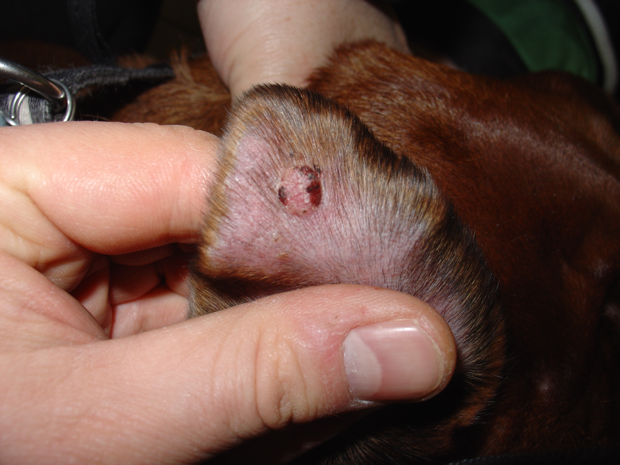
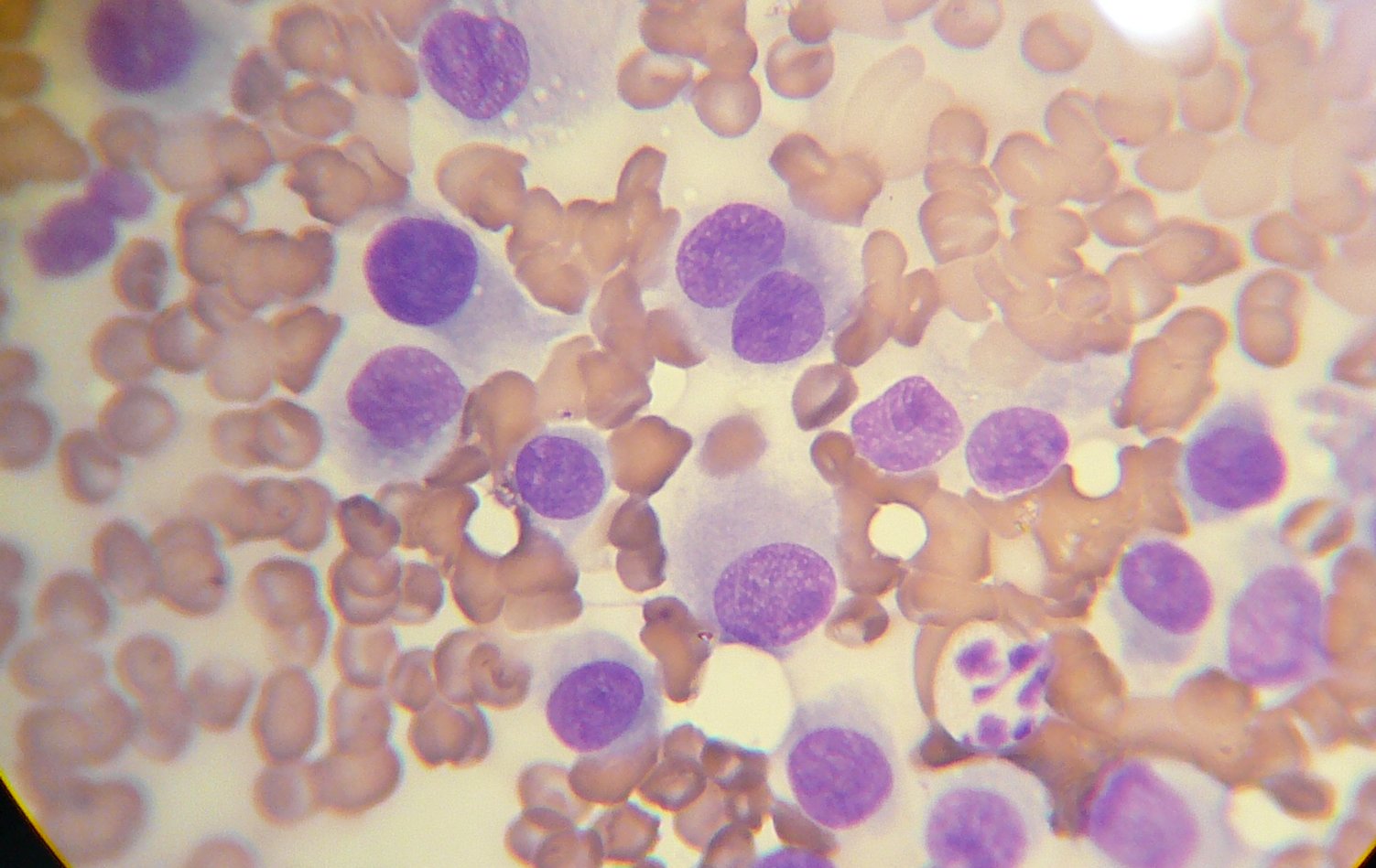
Most commonly histiocytomas are found in young dogs and appear as a small, solitary, hairless lump, although Shar Peis may be predisposed to multiple histiocytomas. They are most commonly found on the head, neck, ears, and limbs, and are usually less than 2.5 cm in diameter. Ulceration of the mass is common. Diagnosis is made through cytology of the mass. Cytology reveals cells with clear to lightly basophilic cytoplasm and round or indented nuclei with fine chromatin and indistinct nucleoli.
Treatment
Most histiocytomas will regress within two or three months. Histiocytomas can regress and disappear completely using antihistamine medications: H1 antagonist is diphenhydramine (Benadryl), and/or the H2 antagonist famotidine (Pepsid). Histiocyte growth can be suppressed using either or both of the H1/H2 antagonists. This can be curative. Surgical removal may be necessary if the tumor does not regress or if it is growing rapidly to a large size. Histiocytomas should never be treated with an intralesional injection of a corticosteroid, as remission relies on recognition of the tumour by the body's immune system which is suppressed by steroids.
References
References
- (1995). "[Dendritic cells in dogs and cats: models of study in human pathology]". Pathol. Biol..
- (2006). "Tumors with Histiocytic Differentiation". The Merck Veterinary Manual.
- (2006). "Langerhans cells arise from monocytes ''in vivo''". Nat. Immunol..
- (1996). "Canine cutaneous histiocytoma is an epidermotropic Langerhans cell histiocytosis that expresses CD1 and specific beta 2-integrin molecules". Am. J. Pathol..
- (2006). "The regression of a canine Langerhans cell tumour is associated with increased expression of IL-2, TNF-alpha, IFN-gamma and iNOS mRNA". Immunology.
- Morrison, Wallace B.. (1998). "Cancer in Dogs and Cats". Williams and Wilkins.
- Cronin, Kim. (Dec 2006). "Deciphering the histiocytic code". Advanstar Communications.
- Affolter, Verena K.. (2004). "Histiocytic Proliferative Diseases in Dogs and Cats". Proceedings of the 29th World Congress of the World Small Animal Veterinary Association.
- Raskin, R.E.. (2006). "Cytology of Neoplasia". Proceedings of the North American Veterinary Conference.
This article was imported from Wikipedia and is available under the Creative Commons Attribution-ShareAlike 4.0 License. Content has been adapted to SurfDoc format. Original contributors can be found on the article history page.
Ask Mako anything about Histiocytoma (dog) — get instant answers, deeper analysis, and related topics.
Research with MakoFree with your Surf account
Create a free account to save articles, ask Mako questions, and organize your research.
Sign up freeThis content may have been generated or modified by AI. CloudSurf Software LLC is not responsible for the accuracy, completeness, or reliability of AI-generated content. Always verify important information from primary sources.
Report