From Surf Wiki (app.surf) — the open knowledge base
HER2
Mammalian protein found in humans
Mammalian protein found in humans
Receptor tyrosine-protein kinase erbB-2 is a protein that normally resides in the membranes of cells and is encoded by the ERBB2 gene. ERBB is abbreviated from erythroblastic oncogene B, a gene originally isolated from the avian genome. The human protein is also frequently referred to as HER2 (human epidermal growth factor receptor 2) or CD340 (cluster of differentiation 340).
HER2 is a member of the human epidermal growth factor receptor (HER/EGFR/ERBB) family. But contrary to other members of the ERBB family, HER2 does not directly bind ligand. HER2 activation results from heterodimerization with another ERBB member or by homodimerization when HER2 concentration are high, for instance in cancer. Amplification or over-expression of this oncogene has been shown to play an important role in the development and progression of certain aggressive types of breast cancer. In recent years the protein has become an important biomarker and target of therapy for approximately 30% of breast cancer patients.
Name
HER2 is named as such due to structural similarities with human epidermal growth factor receptor 1, or HER1*.*
The Neu alias of HER2 derives its name from the parent glioblastoma cell line - a type of neural tumor found in rodents.
ErbB-2 was named for its similarity to avian erythroblastosis oncogene B*, ErbB*; the oncogene later shown to code for epidermal growth factor receptor, EGFR.
Molecular cloning of EGFR discovered that HER2, Neu, and ErbB-2 are all encoded by the same orthologs.
Gene
ERBB2, a known proto-oncogene, is located at the long arm of human chromosome 17 (17q12).
Function
The ErbB family consists of four individual plasma membrane-bound receptor tyrosine kinases. One of which is erbB-2, and the other members being erbB-1, erbB-3 (neuregulin-binding; lacks kinase domain), and erbB-4. All four contain an extracellular ligand binding domain, a transmembrane domain, and an intracellular domain that can interact with a multitude of signaling molecules and exhibit both ligand-dependent and ligand-independent activity. Notably, no ligands for HER2 have yet been identified. HER2 can heterodimerise with any of the other three receptors and is considered to be the preferred dimerisation partner of the other ErbB receptors.
Dimerisation results in the autophosphorylation of tyrosine residues within the cytoplasmic domain of the receptors and initiates a variety of signaling pathways.
Signal transduction
Signaling pathways activated by HER2 include:
- mitogen-activated protein kinase (MAPK)
- phosphoinositide 3-kinase (PI3K/Akt)
- phospholipase C γ
- protein kinase C (PKC)
- Signal transducer and activator of transcription (STAT)
In summary, signaling through the ErbB family of receptors promotes cell proliferation and opposes apoptosis, and therefore must be tightly regulated to prevent uncontrolled cell growth from occurring.
Clinical significance
Cancer
Amplification, also known as the over-expression of the ERBB2 gene, occurs in approximately 15-30% of breast cancers. HER2-positive breast cancers are well established as being associated with increased disease recurrence and a poor prognosis compared with other identifiably genetically distinct breast cancers with other known, or lack thereof, genetic markers that are thought to be associated with other breast cancers; however, drug agents targeting HER2 in breast cancer have significantly and positively altered the otherwise poor prognosis of the historically problematic difficulties associated with HER2-positive breast cancer. Over-expression is also known to occur in ovarian, stomach, adenocarcinoma of the lung and aggressive forms of uterine cancer, such as uterine serous endometrial carcinoma, e.g. HER2 is over-expressed in approximately 7-34% of patients with gastric cancer and in 30% of salivary duct carcinomas.
HER2 is colocalised and most of the time, coamplified with the gene GRB7, which is a proto-oncogene associated with breast, testicular germ cell, gastric, and esophageal tumours.
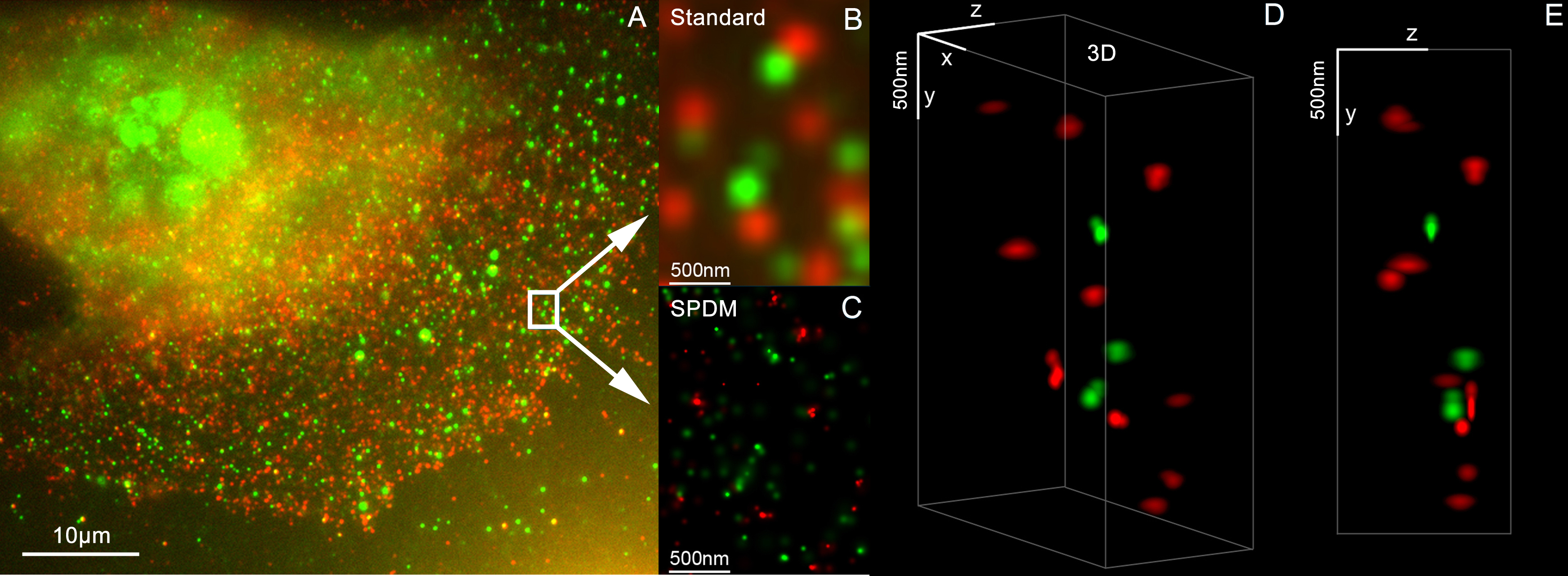
HER2 proteins have been shown to form clusters in cell membranes that may play a role in tumorigenesis.
Evidence has also implicated HER2 signaling in resistance to the EGFR-targeted cancer drug cetuximab.
The high expression of HER2 correlates with better survival in esophageal adenocarcinoma.
The high amplification of HER2 copy number positively contributes to the survival time of gastric cardia adenocarcinoma patients.
Mutations
Furthermore, diverse structural alterations have been identified that cause ligand-independent firing of this receptor, doing so in the absence of receptor over-expression. HER2 is found in a variety of tumours and some of these tumours carry point mutations in the sequence specifying the transmembrane domain of HER2. Substitution of a valine for a glutamic acid or a glutamine in the transmembrane domain can result in the constitutive dimerisation of this protein in the absence of a ligand.
HER2 mutations have been found in non-small-cell lung cancers (NSCLC) and can direct treatment.
As a drug target
HER2 is the target of the monoclonal antibody trastuzumab (marketed as Herceptin). Trastuzumab is effective only in cancers where HER2 is over-expressed. One year of trastuzumab therapy is recommended for all patients with HER2-positive breast cancer who are also receiving chemotherapy. Randomized trials have demonstrated no additional benefit beyond 12 months, whereas 6 months has been shown to be inferior to 12. Trastuzumab is administered intravenously weekly or every 3 weeks.
An important downstream effect of trastuzumab binding to HER2 is an increase in p27, a protein that halts cell proliferation. Another monoclonal antibody, Pertuzumab, which inhibits dimerisation of HER2 and HER3 receptors, was approved by the FDA for use in combination with trastuzumab in June 2012.
As of November 2015, there are a number of ongoing and recently completed clinical trials of novel targeted agents for HER2+ metastatic breast cancer, e.g. margetuximab.
Additionally, NeuVax (Galena Biopharma) is a peptide-based immunotherapy that directs "killer" T cells to target and destroy cancer cells that express HER2. It has entered phase 3 clinical trials.
It has been found that patients with ER+ (estrogen receptor positive)/HER2+ compared with ER-/HER2+ breast cancers may actually benefit more from drugs that inhibit the PI3K/AKT molecular pathway.
Over-expression of HER2 can also be suppressed by the amplification of other genes. Research is currently being conducted to discover which genes may have this desired effect.
The expression of HER2 is regulated by signaling through estrogen receptors. Normally, estradiol and tamoxifen acting through the estrogen receptor down-regulate the expression of HER2. However, when the ratio of the coactivator AIB-3 exceeds that of the corepressor PAX2, the expression of HER2 is upregulated in the presence of tamoxifen, leading to tamoxifen-resistant breast cancer.
Among approved anti-HER2 therapeutics are also tyrosine kinase inhibitors (such as lapatinib, neratinib, and tucatinib) and antibody-drug conjugates (ado-trastuzumab emtansine and trastuzumab deruxtecan).
Diagnostics
HER2 testing is performed on breast biopsy of breast cancer patients to assess prognosis and to determine suitability for trastuzumab therapy. It is important that trastuzumab is restricted to HER2-positive individuals as it is expensive and has been associated with cardiac toxicity. For HER2-positive tumors, the benefits of trastuzumab clearly outweigh the risks.
Tests are usually performed on breast biopsy samples obtained by either fine-needle aspiration, core needle biopsy, vacuum-assisted breast biopsy, or surgical excision.
Immunohistochemistry (IHC) is generally used to measure the amount of HER2 protein present in the sample, with fluorescence in situ hybridisation (FISH) being used on samples that are equivocal in IHC. However, in several locations, FISH is used initially, followed by IHC in equivocal cases.
Immunohistochemistry
By immunohistochemistry, the sample is given a score based on the cell membrane staining pattern.
| title = IHC (ImmunoHistoChemistry) Tests | url = https://www.breastcancer.org/screening-testing/ihc-immunohistochemistry-tests | website=Breastcancer.org | access-date=2019-10-04 }} | url=https://documents.cap.org/documents/algorithim-evaluation-her2.pdf | title=Figure 1. Algorithm for evaluation of human epidermal growth factor receptor 2 (HER2) protein expression by immunohistochemistry (IHC) assay of the invasive component of a breast cancer specimen. | website=College of American Pathologists: Homepage | access-date=2022-09-12}} |
|---|---|---|---|---|---|---|---|
| - | Status | ||||||
| 0 | Either: | HER2 negative | |||||
| (not present) | |||||||
| 1+ | Incomplete membrane staining that is faint or barely perceptible and within 10% of the invasive tumor cells. | ||||||
| 2+ | Weak to moderate complete membrane staining observed in 10% of tumor cells. | Borderline/Equivocal | |||||
| 3+ | Circumferential membrane staining that is complete, intense, and in 10% of tumor cells. | HER2 positive |
Micrographs showing each score: File:HER2 immunohistochemistry with 0 pattern.png|0 File:HER2 immunohistochemistry with 1 plus pattern.png|1+ File:HER2 immunohistochemistry with 2 plus pattern.png|2+ File:HER2 immunohistochemistry with 3 plus pattern.png|3+
Fluorescence ''in situ'' hybridisation
FISH can be used to measure the number of copies of the gene which are present and is thought to be more reliable than immunohistochemistry. It usually uses chromosome enumeration probe 17 (CEP17) to count the amount of chromosomes. Hence, the HER2/CEP17 ratio reflects any amplification of HER2 as compared to the number of chromosomes. The signals of 20 cells are usually counted. File:HER2 FISH with 2 HER2 signals and 3 CEP17 signals.png|This cell displays 2 signals of HER2 (red) and 3 signals of CEP17 (green) File:Counting one or two signals on HER2 FISH.png|Two signals that are closer to each other than the signal diameter count as one. File:HER2 FISH with debris.png|One of these signals is too faint, and is presumably debris. File:HER2 FISH with only one signal type.jpg|Cells with only one type of signal are excluded from the count. File:HER2 FISH with overlapping cells.png|Overlapping cells are also excluded from the count. File:HER2 FISH with a yellow signal.png|A yellow signal counts as one red and one green (which are overlapping) File:Counting HER2 versus CEP17 probes in FISH of HER2 amplified cell.png File:HER2 FISH algorithm.svg|Algorithm for the evaluation of HER2 on fluorescence in situ hybridization (FISH).
| HER2/CEP17 ratio | ≥2.0 | Average HER2 copy number per cell | ≥4.0 |
|---|---|---|---|
| HER2 positive | Additional work-up required | ||
| Additional work-up required | HER2 negative |
If the initial HER2 result is negative for a needle biopsy of a primary breast cancer, a new HER2 test may be performed on the subsequent breast excision.
Serum
The extracellular domain of HER2 can be shed from the surface of tumour cells and enter the circulation. Measurement of serum HER2 by enzyme-linked immunosorbent assay (ELISA) offers a far less invasive method of determining HER2 status than a biopsy and consequently has been extensively investigated. Results so far have suggested that changes in serum HER2 concentrations may be useful in predicting response to trastuzumab therapy. However, its ability to determine eligibility for trastuzumab therapy is less clear.
Interactions
HER2/neu has been shown to interact with:
- CTNNB1
- DLG4
- Erbin
- GRB2
- HSP90AA1
- IL6ST
- MUC1
- PICK1 and
- PIK3R2
- PLCG1 and
- SHC1
References
References
- "ERBB2 erb-b2 receptor tyrosine kinase 2 [Homo sapiens (human)] - Gene - NCBI".
- "ERBB2".
- (2015-01-22). "Noninvasive Molecular Markers in Gynecologic Cancers". CRC Press.
- (2016). "The role of HER2, EGFR, and other receptor tyrosine kinases in breast cancer". Cancer and Metastasis Reviews.
- (December 1985). "Tyrosine kinase receptor with extensive homology to EGF receptor shares chromosomal location with neu oncogene". Science.
- (January 1986). "The neu oncogene encodes an epidermal growth factor receptor-related protein". Nature.
- (March 1986). "Localization of a novel v-erbB-related gene, c-erbB-2, on human chromosome 17 and its amplification in a gastric cancer cell line". Molecular and Cellular Biology.
- (June 2002). "Mechanism of 17-beta-estradiol-induced Erk1/2 activation in breast cancer cells. A role for HER2 AND PKC-delta". The Journal of Biological Chemistry.
- (October 2001). "The characterization of novel, dual ErbB-2/EGFR, tyrosine kinase inhibitors: potential therapy for cancer". Cancer Research.
- (2001). "Update on HER-2 as a target for cancer therapy: intracellular signaling pathways of ErbB2/HER-2 and family members". Breast Cancer Research.
- (November 2009). "Beyond trastuzumab: small molecule tyrosine kinase inhibitors in HER-2-positive breast cancer". The Oncologist.
- (2012). "The HER2 Receptor in Breast Cancer: Pathophysiology, Clinical Use, and New Advances in Therapy". Chemotherapy Research and Practice.
- (October 2005). "The distinctive nature of HER2-positive breast cancers". The New England Journal of Medicine.
- (2007). "Breast Cancer Chemosensitivity".
- (2013). "Robbins basic pathology". Elsevier/Saunders.
- (2013). "Robbins basic pathology". Elsevier/Saunders.
- (August 2008). "Trastuzumab treatment in patients with advanced or recurrent endometrial carcinoma overexpressing HER2/neu". International Journal of Gynaecology and Obstetrics.
- (March 2014). "HER2/neu in Endometrial Cancer: A Promising Therapeutic Target With Diagnostic Challenges". Archives of Pathology & Laboratory Medicine.
- (May 2012). "HER2 testing in gastric cancer: a practical approach". Modern Pathology.
- (March 2011). "Critical appraisal of trastuzumab in treatment of advanced stomach cancer". Cancer Management and Research.
- (June 2015). "Molecular characterization of apocrine salivary duct carcinoma". The American Journal of Surgical Pathology.
- (June 1999). "Activation-dependent clustering of the erbB2 receptor tyrosine kinase detected by scanning near-field optical microscopy". Journal of Cell Science.
- (April 2011). "Analysis of Her2/neu membrane protein clusters in different types of breast cancer cells using localization microscopy". Journal of Microscopy.
- (September 2011). "Activation of ERBB2 signaling causes resistance to the EGFR-directed therapeutic antibody cetuximab". Science Translational Medicine.
- (January 2019). "HER2/neu (ERBB2) expression and gene amplification correlates with better survival in esophageal adenocarcinoma". BMC Cancer.
- (November 2021). "Focal amplifications are associated with chromothripsis events and diverse prognoses in gastric cardia adenocarcinoma". Nature Communications.
- (November 1990). "Correlation of the structure of the transmembrane domain of the neu oncogene-encoded p185 protein with its function". Proceedings of the National Academy of Sciences of the United States of America.
- (June 2013). "Lung cancer that harbors an HER2 mutation: epidemiologic characteristics and therapeutic perspectives". Journal of Clinical Oncology.
- (March 2015). "Systemic targeted therapy for her2-positive early female breast cancer: a systematic review of the evidence for the 2014 Cancer Care Ontario systemic therapy guideline". Current Oncology.
- (2018). "Harrison's Principles of Internal Medicine". McGraw-Hill Education.
- (January 2005). "HER2-targeting antibodies modulate the cyclin-dependent kinase inhibitor p27Kip1 via multiple signaling pathways". Cell Cycle.
- (November 2015). "Human epidermal growth factor receptor 2 positive (HER2+) metastatic breast cancer: how the latest results are improving therapeutic options". Therapeutic Advances in Medical Oncology.
- (June 2009). "Gene expression profiling identifies activated growth factor signaling in poor prognosis (Luminal-B) estrogen receptor positive breast cancer". BMC Medical Genomics.
- (2008-11-13). "Study sheds new light on tamoxifen resistance". Cordis.
- (December 2008). "Regulation of ERBB2 by oestrogen receptor-PAX2 determines response to tamoxifen". Nature.
- (February 2021). "Targeting HER2 expression in cancer: New drugs and new indications". Bosnian Journal of Basic Medical Sciences.
- (August 2007). "Trastuzumab-related cardiotoxicity: calling into question the concept of reversibility". Journal of Clinical Oncology.
- (November 2016). "FISH testing of HER2 immunohistochemistry 1+ invasive breast cancer with unfavorable characteristics". Oncology Letters.
- "IHC (ImmunoHistoChemistry) Tests".
- (2014). "Human Epidermal Growth Factor Receptor 2 (HER2) in Cancers: Overexpression and Therapeutic Implications". Molecular Biology International.
- (January 2020). "Figure 1. Algorithm for evaluation of human epidermal growth factor receptor 2 (HER2) protein expression by immunohistochemistry (IHC) assay of the invasive component of a breast cancer specimen.". Journal of Pathology and Translational Medicine.
- (May 2012). "A gene-protein assay for human epidermal growth factor receptor 2 (HER2): brightfield tricolor visualization of HER2 protein, the HER2 gene, and chromosome 17 centromere (CEN17) in formalin-fixed, paraffin-embedded breast cancer tissue sections". Diagnostic Pathology.
- (2019). "Current Medical Diagnosis & Treatment". McGraw-Hill.
- (July 2018). "Human Epidermal Growth Factor Receptor 2 Testing in Breast Cancer: American Society of Clinical Oncology/College of American Pathologists Clinical Practice Guideline Focused Update". Journal of Clinical Oncology.
- (September 2008). "Serum HER-2/neu and relative resistance to trastuzumab-based therapy in patients with metastatic breast cancer". Cancer.
- (April 2009). "Utility of serum HER2 extracellular domain assessment in clinical decision making: pooled analysis of four trials of trastuzumab in metastatic breast cancer". Journal of Clinical Oncology.
- (June 2002). "ErbB-beta-catenin complexes are associated with human infiltrating ductal breast and murine mammary tumor virus (MMTV)-Wnt-1 and MMTV-c-Neu transgenic carcinomas". The Journal of Biological Chemistry.
- (February 2001). "Geldanamycin abrogates ErbB2 association with proteasome-resistant beta-catenin in melanoma cells, increases beta-catenin-E-cadherin association, and decreases beta-catenin-sensitive transcription". Cancer Research.
- (March 1995). "c-erbB-2 gene product directly associates with beta-catenin and plakoglobin". Biochemical and Biophysical Research Communications.
- (May 2000). "Regulation of neuregulin signaling by PSD-95 interacting with ErbB4 at CNS synapses". Neuron.
- (May 2001). "The ERBB2/HER2 receptor differentially interacts with ERBIN and PICK1 PSD-95/DLG/ZO-1 domain proteins". The Journal of Biological Chemistry.
- (July 2000). "Collective nomenclature for LAP proteins". Nature Cell Biology.
- (January 2003). "Erbin suppresses the MAP kinase pathway". The Journal of Biological Chemistry.
- (2005). "Phosphotyrosine interactome of the ErbB-receptor kinase family". Molecular Systems Biology.
- (December 2001). "Hyaluronan promotes CD44v3-Vav2 interaction with Grb2-p185(HER2) and induces Rac1 and Ras signaling during ovarian tumor cell migration and growth". The Journal of Biological Chemistry.
- (September 1998). "ErbB-1 and ErbB-2 acquire distinct signaling properties dependent upon their dimerization partner". Molecular and Cellular Biology.
- (February 2001). "Sensitivity of mature Erbb2 to geldanamycin is conferred by its kinase domain and is mediated by the chaperone protein Hsp90". The Journal of Biological Chemistry.
- (October 2008). "Quercetin-induced ubiquitination and down-regulation of Her-2/neu". Journal of Cellular Biochemistry.
- (January 2002). "An unexpected biochemical and functional interaction between gp130 and the EGF receptor family in breast cancer cells". Oncogene.
- (August 2003). "Heregulin targets gamma-catenin to the nucleolus by a mechanism dependent on the DF3/MUC1 oncoprotein". Molecular Cancer Research.
- (April 2001). "Transgenic MUC1 interacts with epidermal growth factor receptor and correlates with mitogen-activated protein kinase activation in the mouse mammary gland". The Journal of Biological Chemistry.
- (December 1992). "Expression and characterization of the p85 subunit of the phosphatidylinositol 3-kinase complex and a related p85 beta protein by using the baculovirus expression system". The Biochemical Journal.
- (August 1991). "Oncogenic forms of the neu/HER2 tyrosine kinase are permanently coupled to phospholipase C gamma". The EMBO Journal.
- (December 1991). "Elevated content of the tyrosine kinase substrate phospholipase C-gamma 1 in primary human breast carcinomas". Proceedings of the National Academy of Sciences of the United States of America.
- (March 1999). "A differential requirement for the COOH-terminal region of the epidermal growth factor (EGF) receptor in amphiregulin and EGF mitogenic signaling". The Journal of Biological Chemistry.
This article was imported from Wikipedia and is available under the Creative Commons Attribution-ShareAlike 4.0 License. Content has been adapted to SurfDoc format. Original contributors can be found on the article history page.
Ask Mako anything about HER2 — get instant answers, deeper analysis, and related topics.
Research with MakoFree with your Surf account
Create a free account to save articles, ask Mako questions, and organize your research.
Sign up freeThis content may have been generated or modified by AI. CloudSurf Software LLC is not responsible for the accuracy, completeness, or reliability of AI-generated content. Always verify important information from primary sources.
Report