From Surf Wiki (app.surf) — the open knowledge base
Hemoglobinopathy
Any of various genetic disorders of blood

Any of various genetic disorders of blood
| Field | Value |
|---|---|
| name | Hemoglobinopathy |
| synonyms | Hemoglobinopathies |
| image | File:Sickle cells.jpg |
| caption | Red blood cells from a person with sickle cell disease, illustrating abnormal 'sickle' shaped red blood cells - key characteristic of the disease. |
| symptoms | Chronic anemia |
| complications | Enlarged spleen, iron overload, death |
| onset | During fetal development or very early infancy |
| types | Relatively frequent: sickle cell disease, alpha thalassemia and beta thalassemia |
| causes | Usually inherited |
| diagnosis | Blood smear, ferritin test, hemoglobin electrophoresis, DNA sequencing |
| differential | Iron deficiency anemia |
| prevention | Genetic counselling of potential parents, termination of pregnancy |
| treatment | Blood transfusion, iron chelation, hematopoietic stem cell transplant |
Hemoglobinopathy is the medical term for a group of inherited blood disorders involving the hemoglobin, the major protein of red blood cells. They are generally single-gene disorders and, in most cases, they are inherited as autosomal recessive traits.
There are two main groups: abnormal structural hemoglobin variants caused by mutations in the hemoglobin genes, and the thalassemias, which are caused by an underproduction of otherwise normal hemoglobin molecules. The main structural hemoglobin variants are HbS, HbE and HbC. The main types of thalassemia are alpha-thalassemia and beta thalassemia.
Hemoglobin functions
Hemoglobin is a protein containing iron that facilitates the transportation of oxygen in red blood cells. Hemoglobin in the blood carries oxygen from the lungs to the other tissues of the body, where it releases the oxygen to enable aerobic respiration which powers the metabolism. Normal levels of hemoglobin vary according to sex and age in the range 9.5 to 17.2 grams of hemoglobin in every deciliter of blood.
Hemoglobin also transports other gases. It carries off some of the body's respiratory carbon dioxide (about 20–25% of the total) as carbaminohemoglobin, in which CO2 binds to the heme protein. The molecule also carries the important regulatory molecule nitric oxide bound to a thiol group in the globin protein, releasing it at the same time as oxygen.
Hemoglobin structural biology
Normal human hemoglobins are tetrameric proteins composed of two pairs of globin chains, each of which contains one alpha-like (α) globin and one beta-like (β) globin. Each globin chain is associated with an iron-containing heme moiety. Throughout life, the synthesis of the α and the β chains is balanced so that their ratio is relatively constant and there is no excess of either type.
The specific α and β chains that are incorporated into Hb are highly regulated during development:
- Embryonic Hb are expressed as early as four to six weeks of embryogenesis and disappear around the eighth week of gestation as they are replaced by fetal Hb. Embryonic Hbs include:
- Hb Gower-1, composed of two ζ (zeta) globins and two ε (epsilon) globins, i.e., ζ2ε2
- Hb Gower-2, composed of two α globins and two ε globins (α2ε2)
- Hb Portland, composed of two ζ globins and two γ (gamma) globins (ζ2γ2)
- Fetal Hb (HbF) is produced from approximately eight weeks of gestation through birth and constitutes approximately 80 percent of Hb in the full-term neonate. It declines during the first few months of life and, in the normal state, constitutes 2γ2).
- Adult Hb (HbA) is the predominant Hb in children by six months of age and onward; it constitutes 96-97% of total Hb in individuals without a hemoglobinopathy. It is composed of two α globins and two β globins (α2β2).
- HbA2 is a minor adult Hb that normally accounts for approximately 2.5–3.5% of total Hb from six months of age onward. It is composed of two α globins and two δ (delta) globins (α2δ2).
Classification of hemoglobinopathies
A) Qualitative
Structural abnormalities
Hemoglobin structural variants manifest a change in the structure of the Hb molecule. The majority of hemoglobin variants do not cause disease and are most commonly discovered either incidentally or through newborn screening. Hb variants can usually be detected by protein-based assay methods such as electrophoresis, isoelectric focusing, or high-performance liquid chromatography. Diagnosis is commonly confirmed by DNA sequencing.
The hemoglobin structural variants can be broadly classified as follows:
- Sickle cell disorders, which are the most prevalent form of hemoglobinopathy. Sickle hemoglobin (HbS) is prone to polymerize when deoxygenated, precipitating within the red blood cell. This damages the RBC membrane resulting in its premature destruction and consequent anemia.
- Unstable hemoglobin variants are mutations that cause the hemoglobin molecule to precipitate, spontaneously or upon oxidative stress, resulting in hemolytic anemia. Precipitated, denatured hemoglobin can attach to the inner layer of the plasma membrane of the red blood cell (RBC) forming Heinz bodies, leading to premature destruction of the RBC and anemia.
- Change in oxygen affinity. High or low oxygen affinity hemoglobin molecules are more likely than normal to adopt the relaxed (R, oxy) state or the tense (T, deoxy) state, respectively. High oxygen affinity variants (R state) cause polycythemia (e.g., Hb Chesapeake, Hb Montefiore). Low oxygen affinity variants can cause cyanosis (e.g., Hb Kansas, Hb Beth Israel).
Chemical abnormalities
Methemoglobinemia is a condition caused by elevated levels of methemoglobin in the blood. Methaemoglobin is a form of hemoglobin that contains the ferric [Fe3+] form of iron, instead of the ferrous [Fe2+] form . Methemoglobin cannot bind oxygen, which means it cannot carry oxygen to tissues. In human blood a trace amount of methemoglobin is normally produced spontaneously; the enzyme methemoglobin reductase is responsible for converting methemoglobin back to hemoglobin. Methemoglobinemia can be hereditary but more commonly occurs as a side effect of certain medications or by abuse of recreational drugs.
B) Quantitative
Production abnormalities

The severity of alpha thalassemia depends on how many of the four genes that code for alpha globin are faulty. In the fetus, a deficiency of alpha globin results in the production of Hemoglobin Barts - a dysfunctional hemoglobin that consists of four gamma globins. In this situation, a fetus will develop hydrops fetalis and normally die before or shortly after birth. In adults alpha thalassemia manifests as HbH disease. In this, excess beta-globin forms β4-tetramers, which accumulate and precipitate in red blood cells, damaging their membranes. Damaged RBCs are removed by the spleen resulting in moderate to severe anemia.
In beta thalassemia, reduced production of beta globin, combined with a normal synthesis of alpha globin, results in an accumulation of excess unmatched alpha globin. This precipitates in the red cell precursors in the bone marrow, triggering their premature destruction. Anemia in beta thalassemia results from a combination of ineffective production of RBCs, peripheral hemolysis, and an overall reduction in hemoglobin synthesis.
Combination hemoglobinopathies
A combination hemoglobinopathy occurs when someone inherits two different abnormal hemoglobin genes. If these are different versions of the same gene, one having been inherited from each parent it is an example of compound heterozygosity.
Both alpha- and beta- thalassemia can coexist with other hemoglobinopathies. Combinations involving alpha thalassemia are generally benign.
Some examples of clinically significant combinations involving beta thalassemia include:
-
Hemoglobin C/ beta thalassemia: common in Mediterranean and African populations generally results in a moderate form of anemia with splenomegaly.
-
Hemoglobin D/ beta thalassemia: common in the northwestern parts of India and Pakistan (Punjab region).
-
Hemoglobin E/ beta thalassemia: common in Cambodia, Thailand, and parts of India, it is clinically similar to β thalassemia major or β thalassemia intermedia.
-
Hemoglobin S/ beta thalassemia: common in African and Mediterranean populations, it is clinically similar to sickle-cell anemia.
-
Delta-beta thalassemia is a rare form of thalassemia in which there is a reduced production of both the delta and beta globins. It is generally asymptomatic.
There are two clinically significant combinations involving the sickle cell gene:
- Hemoglobin S/ beta thalassemia: (see above).
- Hemoglobin S/ hemoglobin C (Hemoglobin SC disease) occurs when an individual inherits one gene for hemoglobin S (sickle cell) and one gene for hemoglobin C, The symptoms are very similar to sickle cell disease.
Hemoglobin variants
Hemoglobin variants are not necessarily pathological. For example, Hb Lepore-Boston and G-Waimanalo are two variants which are non-pathological. There are in excess of 1,000 known hemoglobin variants. A research database of hemoglobin variants is maintained by Penn State University. A few of these variants are listed below.
Normal hemoglobins
Source: ; Embryonic
- HbE Gower 1 (ζ2ε2) present in the normal embryo.
- HbE Gower 2 (α2ε2) present in the normal embryo.
- HbE Portland I (ζ2γ2) present in the normal embryo.
; Fetal
- HbF/Fetal (α2γ2) dominating during pregnancy and reducing close to zero a few weeks after birth
- HbA (α2β2) Adult hemoglobin, present in small quantities during pregnancy
; Adult
- HbA (α2β2) comprising approximately 97% of adult hemoglobin
- HbA2 (α2δ2) comprising approximately 3% of adult hemoglobin
- HbF/Fetal (α2γ2) dominating during pregnancy and reducing close to zero after birth
Relatively common abnormal hemoglobins
Source:
- HbS (α2βS2) causing sickle cell disease
- HbC (α2βC2) causing mild anemia if homozygous
- HbE (α2βE2) causing mild anemia if homozygous
- HbD causing mild anemia if homozygous
- HbH formed from 4 beta globins in severe alpha thalassemia causing severe anemia
Evolutionary advantage
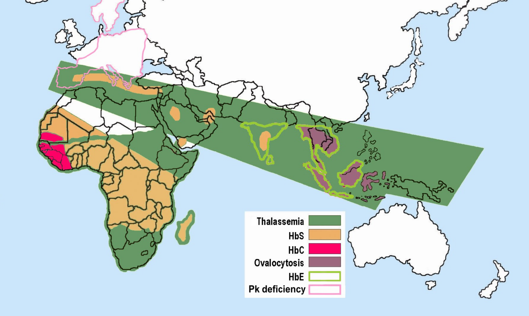
Main article: Malaria resistance
Some hemoglobinopathies seem to have given an evolutionary benefit, especially to heterozygotes, in areas where malaria is endemic. Malaria parasites infect red blood cells, but subtly disturb normal cellular function and subvert the immune response. A number of mechanisms have been proposed to explain the increased chance of survival for the carrier of an abnormal hemoglobin trait.
References
References
- CDC. (2019-02-08). "Hemoglobinopathies Research".
- (2001). "Inherited haemoglobin disorders: An increasing global health problem". Bulletin of the World Health Organization.
- Shakeel, Hassan. (25 March 2023). "Thalassaemia — Knowledge Hub".
- "Hemoglobinopathies and Thalassemia".
- (1993). "Human Biology and Health". Prentice Hall.
- "Hemoglobin: MedlinePlus Medical Encyclopedia".
- Patton, Kevin T.. (2015-02-10). "Anatomy and Physiology". Elsevier Health Sciences.
- (1998). "Respiratory Function of Hemoglobin". New England Journal of Medicine.
- Weatherall DJ. The New Genetics and Clinical Practice, Oxford University Press, Oxford 1991.
- (2014-11-01). "Flicking the switch: adult hemoglobin expression in erythroid cells derived from cord blood and human induced pluripotent stem cells". Haematologica.
- Huisman TH. The structure and function of normal and abnormal haemoglobins. In: Baillière's Clinical Haematology, Higgs DR, Weatherall DJ (Eds), W.B. Saunders, London 1993. p.1.
- Natarajan K, Townes TM, Kutlar A. Disorders of hemoglobin structure: sickle cell anemia and related abnormalities. In: Williams Hematology, 8th ed, Kaushansky K, Lichtman MA, Beutler E, et al. (Eds), McGraw-Hill, 2010. p.ch.48.
- (November 2008). "Hemoglobin research and the origins of molecular medicine". Blood.
- (17 April 2002). "Hemoglobinopathies".
- "Hemoglobin Electrophoresis: MedlinePlus Medical Test".
- Garfin, David E.. (1990). "[35] Isoelectric focusing". Academic Press.
- (2021-05-05). "Techniques for the Detection of Sickle Cell Disease: A Review". Micromachines.
- Forget, Bernard G.. (2013-02-01). "Classification of the Disorders of Hemoglobin". Cold Spring Harbor Perspectives in Medicine.
- (1990). "Sickle Cell Hemoglobin Polymerization". Advances in Protein Chemistry.
- (1995). "Severe hemolytic anemia associated with the homozygous state for an unstable hemoglobin variant (Hb Bushwick)". Blood.
- (2009). "Identification of high oxygen affinity hemoglobin variants in the investigation of patients with erythrocytosis". Haematologica.
- (January 2019). "StatPearls [Internet].". StatPearls Publishing.
- (June 2014). "Human cytochrome b5 reductase: structure, function, and potential applications". Critical Reviews in Biotechnology.
- "Methemoglobinemia (MetHb): Symptoms, Causes & Treatment".
- Dasgupta, Amitava. (2014-01-01). "Chapter 21 - Hemoglobinopathy". Elsevier.
- "Pathophysiology of alpha thalassemia".
- (2009-01-01). "Hb H disease: clinical course and disease modifiers". Hematology.
- Thein, Swee Lay. (2005-01-01). "Pathophysiology of β Thalassemia—A Guide to Molecular Therapies". Hematology.
- (2021-12-31). "Shared molecular basis, diagnosis, and co-inheritance of alpha and beta thalassemia". Blood Research.
- (2006). "Co-inheritance of α+-thalassaemia and sickle trait results in specific effects on haematological parameters". British Journal of Haematology.
- (February 2011). "Hemoglobin C".
- (March 2015). "Hemoglobin D-Punjab: origin, distribution and laboratory diagnosis". Revista Brasileira de Hematologia e Hemoterapia.
- (May 2008). "Studies in haemoglobin E beta-thalassaemia". British Journal of Haematology.
- Gerber, Gloria F.. (April 2024). "Hemoglobin S–Beta-Thalassemia Disease - Hematology and Oncology".
- (2005). "Textbook Of Practical Physiology - 2Nd Edn.". Orient Blackswan.
- Pitone, Melanie L.. "Hemobglobin SC Disease (for Parents)".
- DiGeorge, Nicholas W.. (2014-03-01). "Fitness for Duty: Two Cases of Rare Hemoglobin Variants in U.S. Navy Recruits". Military Medicine.
- (6 July 2018). "Understanding haemoglobinopathies".
- (December 2024). "A Database of Human Hemoglobin Variants and Thalassemia mutations".
- Manning, Lois R.. (2007). "Human embryonic, fetal, and adult hemoglobins have different subunit interface strengths. Correlation with lifespan in the red cell". Protein Science.
- (2013-05-16). "Hemoglobinopathies: Slicing the Gordian Knot of Plasmodium falciparum Malaria Pathogenesis". PLOS Pathogens.
This article was imported from Wikipedia and is available under the Creative Commons Attribution-ShareAlike 4.0 License. Content has been adapted to SurfDoc format. Original contributors can be found on the article history page.
Ask Mako anything about Hemoglobinopathy — get instant answers, deeper analysis, and related topics.
Research with MakoFree with your Surf account
Create a free account to save articles, ask Mako questions, and organize your research.
Sign up freeThis content may have been generated or modified by AI. CloudSurf Software LLC is not responsible for the accuracy, completeness, or reliability of AI-generated content. Always verify important information from primary sources.
Report