From Surf Wiki (app.surf) — the open knowledge base
Hamstring
Any of the three muscles in the thigh
Any of the three muscles in the thigh
| Field | Value |
|---|---|
| Name | Hamstring |
| Image | Hamstrings.gif |
| Caption | Rotating view of the hamstring muscles |
| Origin | tuberosity of the ischium, linea aspera |
| Insertion | tibia, fibula |
| Blood | inferior gluteal artery, profunda femoris artery |
| Nerve | sciatic nerve (tibial nerve and common fibular nerve) |
| Action | flexion of knee, extension of hip |
| Antagonist | Rectus femoris muscle |
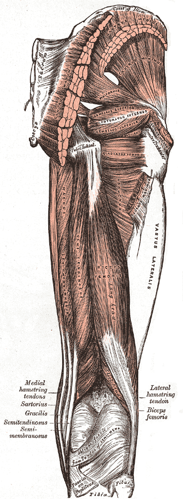
A hamstring () is any one of the three posterior thigh muscles in human anatomy between the hip and the knee: from medial to lateral, the semimembranosus, semitendinosus and biceps femoris.
Etymology
The word "ham" is derived from the Old English “ham” or “hom” meaning the hollow or bend of the knee, from a Germanic base where it meant "crooked". It gained the meaning of the leg of an animal around the 15th century. String refers to tendons, and thus the hamstrings' string-like tendons felt on either side of the back of the knee.
Criteria
The common criteria of any hamstring muscles are:
- Muscles should originate from ischial tuberosity.
- Muscles should be inserted over the knee joint, in the tibia or in the fibula.
- Muscles will be innervated by the tibial branch of the sciatic nerve.
- Muscle will participate in flexion of the knee joint and extension of the hip joint.
Those muscles which fulfill all of the four criteria are called true hamstrings.
The adductor magnus reaches only up to the adductor tubercle of the femur, but it is included amongst the hamstrings because the tibial collateral ligament of the knee joint morphologically is the degenerated tendon of this muscle. The ligament is attached to the medial epicondyle, two millimeters from the adductor tubercle.
Structure
The three muscles of the posterior thigh (semitendinosus, semimembranosus, biceps femoris) flex (bend) the knee, while all but the biceps femoris extend (straighten) the hip. The three 'true' hamstrings cross both the hip and the knee joint and are therefore involved in knee flexion and hip extension. The short head of the biceps femoris crosses only one joint (knee) and is therefore not involved in hip extension. With its divergent origin and innervation, it is sometimes excluded from the 'hamstring' characterization.
| biceps femoris - short head | linea aspera and lateral supracondylar line of femur | lateral side of the head of the fibula (common tendon with the long head) | common peroneal |
|---|
A portion of the adductor magnus is sometimes considered a part of the hamstrings.
Function
The hamstrings cross and act upon two joints – the hip and the knee – and as such they are termed biarticular muscles. The hamstrings contract when the knee is bent or when the hips are extended, and they lengthen when the knee is extended.
Semitendinosus and semimembranosus extend the hip when the trunk is fixed; they also flex the knee and medially (inwardly) rotate the lower leg when the knee is bent.
The long head of the biceps femoris extends the hip, as when beginning to walk; both short and long heads flex the knee and laterally (outwardly) rotate the lower leg when the knee is bent.
The hamstrings play a crucial role in many daily activities such as walking, running, jumping, and controlling some movement in the gluteus. In walking, they are most important as an antagonist to the quadriceps in the deceleration of knee extension.
Clinical significance
Sports running injuries
A common running injury in several sports, excessive stretch of a hamstring results from extensive hip flexion while the knee is extended. During sprinting, a hamstring injury may occur from excessive muscle strain during eccentric contraction late in the leg swing phase. The overall incidence of a hamstring injury in sports and professional dancers is about two per 1000 hours of performance. In some sports, a hamstring injury occurs at the incidence of 19% of all sports injuries, and results in an average time loss from competition of 24 days.
Imaging


Imaging the hamstring muscles is usually performed with an ultrasound and/or MRI. The biceps femoris is most commonly injured, followed by semitendinosus. Semimembranosus injury is rare. Imaging is useful in differentiating the grade of strain, especially if the muscle is completely torn. In this setting, the level and degree of retraction can be determined, serving as a useful roadmap prior to any surgery. Those with a hamstring strain of greater than 60 mm in length have a greater risk of recurrence.
Use in surgery
The distal semitendinosus tendon is one of the tendons that can be used in the surgical procedure ACL reconstruction. In this procedure, a piece of it is used to replace the anterior cruciate ligament (ACL). The ACL is one of the four major ligaments in the knee, which also include the posterior cruciate ligament (PCL), medial collateral ligament (MCL), and lateral collateral ligament (LCL).
References
References
- "University of Glasgow - Schools - School of Life Sciences".
- "Biceps Femoris - Short Head — Musculoskeletal Radiology — UW Radiology". Rad.washington.edu.
- Mayo Clinic Staff. (3 Oct 2015). "Hamstring injury". Mayo clinic.
- (2020-09-29). "The mechanism of hamstring injuries – a systematic review". BMC Musculoskeletal Disorders.
- Brown, Lesley, ed. (2007). Shorter Oxford English Dictionary II (Sixth ed.). Oxford: Oxford University press. p. 3611.
- "Online Etymology Dictionary". Etymonline.com.
- {{NormanAnatomy. postthigh
- (2019-05-22). "Late swing or early stance? A narrative review of hamstring injury mechanisms during high-speed running". Scandinavian Journal of Medicine and Science in Sports.
- (2003). "Evaluation of the hamstring muscle complex following acute injury". Skeletal Radiol..
- (2008). "Rupture of the conjoint tendon at the proximal musculotendinous junction of the biceps femoris long head: a case report.". Knee Surg Sports Traumatol Arthrosc.
- (2007). "Magnetic resonance imaging parameters for assessing risk of recurrent hamstring injuries in elite athletes.". Am J Sports Med.
This article was imported from Wikipedia and is available under the Creative Commons Attribution-ShareAlike 4.0 License. Content has been adapted to SurfDoc format. Original contributors can be found on the article history page.
Ask Mako anything about Hamstring — get instant answers, deeper analysis, and related topics.
Research with MakoFree with your Surf account
Create a free account to save articles, ask Mako questions, and organize your research.
Sign up freeThis content may have been generated or modified by AI. CloudSurf Software LLC is not responsible for the accuracy, completeness, or reliability of AI-generated content. Always verify important information from primary sources.
Report