From Surf Wiki (app.surf) — the open knowledge base
Glia
Support-cells in the nervous system

Support-cells in the nervous system
| Field | Value |
|---|---|
| Name | Glia |
| Image | Glial Cell Types.png |
| Caption | Illustration of the four different types of glial cells found in the central nervous system: ependymal cells (light pink), astrocytes (green), microglial cells (dark red) and oligodendrocytes (light blue) |
| Precursor | Neuroectoderm for macroglia, and hematopoietic stem cells for microglia |
| System | Nervous system |
Glia, also called glial cells (gliocytes) or neuroglia, are non-neuronal cells in the central nervous system (the brain and the spinal cord) and in the peripheral nervous system that do not produce electrical impulses. The neuroglia make up more than one half the volume of neural tissue in the human body. They contribute to the maintenance of homeostasis, help form myelin, and provide support and protection for neurons. In the central nervous system, glial cells include oligodendrocytes (that produce myelin), astrocytes, ependymal cells and microglia, and in the peripheral nervous system they include Schwann cells (that produce myelin), and satellite cells.
Function
Glia have four main functions:
- to structurally support neurons, holding them in place
- to supply nutrients and oxygen to neurons
- to insulate one neuron from another
- to destroy pathogens and remove dead neurons. They also play a role in neurotransmission and synaptic connections, and in physiological processes such as breathing. While glia were thought to outnumber neurons by a ratio of 10:1, studies using newer methods and reappraisal of historical quantitative evidence suggests an overall ratio of less than 1:1, with substantial variation between different brain tissues.
Glial cells have far more cellular diversity and functions than neurons, and can respond to and manipulate neurotransmission in multiple ways. Additionally, they can affect both the preservation and consolidation of memories.
Glia were discovered in 1856, by the pathologist Rudolf Virchow in his search for a "connective tissue" in the brain. The term derives from Greek γλία and γλοία "glue" ( or ), and suggests the original impression that they were the glue of the nervous system.
Types

.jpg)
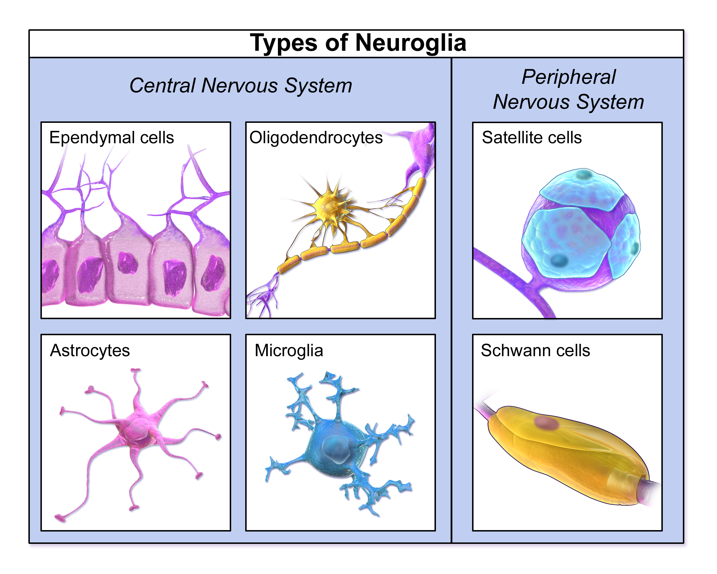
Macroglia
Derived from ectodermal tissue.
| Location | Name | Description |
|---|---|---|
| CNS | Astrocytes | |
| CNS | Oligodendrocytes | |
| CNS | Ependymal cells | |
| CNS | Radial glia | |
| PNS | Schwann cells | |
| PNS | Satellite cells | |
| PNS | Enteric glial cells |
Microglia
Main article: Microglia
Microglia are specialized macrophages capable of phagocytosis that protect neurons of the central nervous system. They are derived from the earliest wave of mononuclear cells that originate in the blood islands of the yolk sac early in development, and colonize the brain shortly after the neural precursors begin to differentiate.
These cells are found in all regions of the brain and spinal cord. Microglial cells are small relative to macroglial cells, with changing shapes and oblong nuclei. They are mobile within the brain and multiply when the brain is damaged. In the healthy central nervous system, microglia processes constantly sample all aspects of their environment (neurons, macroglia and blood vessels). In a healthy brain, microglia direct the immune response to brain damage and play an important role in the inflammation that accompanies the damage. A number of diseases and disorders are associated with deficient microglia, such as Alzheimer's disease, Parkinson's disease and ALS.
Other
Pituicytes from the posterior pituitary are glial cells with characteristics in common to astrocytes. Tanycytes in the median eminence of the hypothalamus are a type of ependymal cell that descend from radial glia and line the base of the third ventricle. Drosophila melanogaster, the fruit fly, contains numerous glial types that are functionally similar to mammalian glia but are nonetheless classified differently.
Total number
In general, neuroglial cells are smaller than neurons. There are approximately 85 billion glial cells in the human brain, about the same number as neurons. Glial cells make up about half the total volume of the brain and spinal cord. The glia to neuron-ratio varies from one part of the brain to another. The glia to neuron-ratio in the cerebral cortex is 3.72 (60.84 billion glia (72%); 16.34 billion neurons), while that of the cerebellum is only 0.23 (16.04 billion glia; 69.03 billion neurons). The ratio in the cerebral cortex gray matter is 1.48, with 3.76 for the gray and white matter combined. The ratio of the basal ganglia, diencephalon and brainstem combined is 11.35.
The total number of glial cells in the human brain is distributed into the different types with oligodendrocytes being the most frequent (45–75%), followed by astrocytes (19–40%) and microglia (about 10% or less).
Development

Main article: Gliogenesis
Most glia are derived from ectodermal tissue of the developing embryo, in particular the neural tube and crest. The exception is microglia, which are derived from hematopoietic stem cells. In the adult, microglia are largely a self-renewing population and are distinct from macrophages and monocytes, which infiltrate an injured and diseased CNS.
In the central nervous system, glia develop from the ventricular zone of the neural tube. These glia include the oligodendrocytes, ependymal cells, and astrocytes. In the peripheral nervous system, glia derive from the neural crest. These PNS glia include Schwann cells in nerves and satellite glial cells in ganglia.
Capacity to divide
Glia retain the ability to undergo cell divisions in adulthood, whereas most neurons cannot. The view is based on the general inability of the mature nervous system to replace neurons after an injury, such as a stroke or trauma, where often there is a substantial proliferation of glia, or gliosis, near or at the site of damage. However, detailed studies have found no evidence that 'mature' glia, such as astrocytes or oligodendrocytes, retain mitotic capacity. Only the resident oligodendrocyte precursor cells seem to keep this ability once the nervous system matures.
Glial cells are known to be capable of mitosis. By contrast, scientific understanding of whether neurons are permanently post-mitotic, or capable of mitosis, is still developing. In the past, glia had been considered to lack certain features of neurons. For example, glial cells were not believed to have chemical synapses or to release transmitters. They were considered to be the passive bystanders of neural transmission. However, recent studies have shown this to not be entirely true.
Functions
Some glial cells function primarily as the physical support for neurons. Others provide nutrients to neurons and regulate the extracellular fluid of the brain, especially surrounding neurons and their synapses. During early embryogenesis, glial cells direct the migration of neurons and produce molecules that modify the growth of axons and dendrites. Some glial cells display regional diversity in the CNS and their functions may vary between the CNS regions.
Neuron repair and development
Glia are crucial in the development of the nervous system and in processes such as synaptic plasticity and synaptogenesis. Glia have a role in the regulation of repair of neurons after injury. In the central nervous system (CNS), glia suppress repair. Glial cells known as astrocytes enlarge and proliferate to form a scar and produce inhibitory molecules that inhibit regrowth of a damaged or severed axon. In the peripheral nervous system (PNS), glial cells known as Schwann cells (or also as neuri-lemmocytes) promote repair. After axonal injury, Schwann cells regress to an earlier developmental state to encourage regrowth of the axon. This difference between the CNS and the PNS, raises hopes for the regeneration of nervous tissue in the CNS. For example, a spinal cord may be able to be repaired following injury or severance.
Myelin sheath creation
Oligodendrocytes are found in the CNS and resemble an octopus: they have a bulbous cell body with up to fifteen arm-like processes. Each process reaches out to an axon and spirals around it, creating a myelin sheath. The myelin sheath insulates the nerve fiber from the extracellular fluid and speeds up signal conduction along the nerve fiber. In the peripheral nervous system, Schwann cells are responsible for myelin production. These cells envelop nerve fibers of the PNS by winding repeatedly around them. This process creates a myelin sheath, which not only aids in conductivity but also assists in the regeneration of damaged fibers.
Neurotransmission
Astrocytes are crucial participants in the tripartite synapse. They have several crucial functions, including clearance of neurotransmitters from within the synaptic cleft, which aids in distinguishing between separate action potentials and prevents toxic build-up of certain neurotransmitters such as glutamate, which would otherwise lead to excitotoxicity. Furthermore, astrocytes release gliotransmitters such as glutamate, ATP, and D-serine in response to stimulation.
Clinical significance

While glial cells in the PNS frequently assist in regeneration of lost neural functioning, loss of neurons in the CNS does not result in a similar reaction from neuroglia. In the CNS, regrowth will only happen if the trauma was mild, and not severe. When severe trauma presents itself, the survival of the remaining neurons becomes the optimal solution. However, some studies investigating the role of glial cells in Alzheimer's disease are beginning to contradict the usefulness of this feature, and even claim it can "exacerbate" the disease. In addition to affecting the potential repair of neurons in Alzheimer's disease, scarring and inflammation from glial cells have been further implicated in the degeneration of neurons caused by amyotrophic lateral sclerosis.
In addition to neurodegenerative diseases, a wide range of harmful exposure, such as hypoxia, or physical trauma, can lead to the result of physical damage to the CNS. Generally, when damage occurs to the CNS, glial cells cause apoptosis among the surrounding cellular bodies. Then, there is a large amount of microglial activity, which results in inflammation, and, finally, there is a heavy release of growth inhibiting molecules.
History
Although glial cells and neurons were probably first observed at the same time in the early 19th century, unlike neurons whose morphological and physiological properties were directly observable for the first investigators of the nervous system, glial cells had been considered to be merely "glue" that held neurons together until the mid-20th century.
Glia were first described in 1856 by the pathologist Rudolf Virchow in a comment to his 1846 publication on connective tissue. A more detailed description of glial cells was provided in the 1858 book 'Cellular Pathology' by the same author.
When markers for different types of cells were analyzed, Albert Einstein's brain was discovered to contain significantly more glia than normal brains in the left angular gyrus, an area thought to be responsible for mathematical processing and language. However, out of the total of 28 statistical comparisons between Einstein's brain and the control brains, finding one statistically significant result is not surprising, and the claim that Einstein's brain is different is not scientific (cf. multiple comparisons problem).
Not only does the ratio of glia to neurons increase through evolution, but so does the size of the glia. Astroglial cells in human brains have a volume 27 times greater than in mouse brains.
These important scientific findings may begin to shift the neurocentric perspective into a more holistic view of the brain which encompasses the glial cells as well. For the majority of the twentieth century, scientists had disregarded glial cells as mere physical scaffolds for neurons. Recent publications have proposed that the number of glial cells in the brain is correlated with the intelligence of a species. Moreover, evidences are demonstrating the active role of glia, in particular astroglia, in cognitive processes like learning and memory.
References
Bibliography
- Kettenmann and Ransom, Neuroglia, Oxford University Press, 2012, |http://ukcatalogue.oup.com/product/9780199794591.do#.UVcswaD3Ay4|
References
- (October 2014). "Glial Biology in Learning and Cognition". The Neuroscientist.
- (August 1980). "Glial cells in the enteric nervous system contain glial fibrillary acidic protein". Nature.
- (July 2008). "D-amino acids in the brain: D-serine in neurotransmission and neurodegeneration". The FEBS Journal.
- Swaminathan, Nikhil. (Jan–Feb 2011). "Glia – the other brain cells". Discover.
- (July 2010). "Astrocytes control breathing through pH-dependent release of ATP". Science.
- (2017). "D-serine released by astrocytes in brainstem regulates breathing response to CO2 levels.". Nat Commun.
- von Bartheld, Christopher S.. (November 2018). "Myths and truths about the cellular composition of the human brain: A review of influential concepts". Journal of Chemical Neuroanatomy.
- "Classic Papers". Max Delbrueck Center für Molekulare Medizin (MDC) Berlin-Buch.
- {{LSJ. gloi/a. γλοία, {{LSJ. gli/a. γλία. ref.
- Swaminathan, N. (2008). "Brain-scan mystery solved". Scientific American Mind.
- Torres A. (2012). "Extracellular Ca2+ Acts as a Mediator of Communication from Neurons to Glia". Science Signaling.
- (2009-10-27). "The Root of Thought: What do Glial Cells Do?".
- (April 2001). "Biology of oligodendrocyte and myelin in the mammalian central nervous system". Physiological Reviews.
- (January 1999). "Identification of a neural stem cell in the adult mammalian central nervous system". Cell.
- Newman EA. (October 2003). "New roles for astrocytes: regulation of synaptic transmission". Trends in Neurosciences.
- (May 2002). "Radial glia: multi-purpose cells for vertebrate brain development". Trends in Neurosciences.
- (September 2005). "The origin and development of glial cells in peripheral nerves". Nature Reviews. Neuroscience.
- Hanani, M. "Satellite glial cells in sensory ganglia: from form to function". ''Brain Res. Rev.'' 48:457–76, 2005
- (December 2008). "Evidence for a role of connexin 43 in trigeminal pain using RNA interference in vivo". Journal of Neurophysiology.
- (July 2007). "Enteric glial cells: new players in gastrointestinal motility?". Laboratory Investigation.
- Brodal, 2010: [https://books.google.com/books?id=iJjI6yDNmr8C&pg=PA19 p. 19]
- "Never-resting microglia: physiological roles in the healthy brain and pathological implications". A Sierra, ME Tremblay, H Wake – 2015 – [https://books.google.com/books?id=GocSBwAAQBAJ&dq=origin+of+microglia&pg=PA6 books.google.com]
- (April 1999). "Morphological plasticity and rearrangement of cytoskeletons in pituicytes cultured from adult rat neurohypophysis.". Neuroscience Research.
- (2005). "Hypothalamic Tanycytes: A Key Component of Brain–Endocrine Interaction".
- Freeman, Marc R.. (2015-02-26). "DrosophilaCentral Nervous System Glia". Cold Spring Harbor Perspectives in Biology.
- (2016-12-15). "The search for true numbers of neurons and glial cells in the human brain: A review of 150 years of cell counting". The Journal of Comparative Neurology.
- (April 2009). "Equal numbers of neuronal and nonneuronal cells make the human brain an isometrically scaled-up primate brain". The Journal of Comparative Neurology.
- (May 2007). "Cell cycle regulation in the postmitotic neuron: oxymoron or new biology?". Nature Reviews. Neuroscience.
- (April 1983). "Neuronal production, migration, and differentiation in a vocal control nucleus of the adult female canary brain". Proceedings of the National Academy of Sciences of the United States of America.
- (November 1998). "Neurogenesis in the adult human hippocampus". Nature Medicine.
- (April 1999). "Hippocampal neurogenesis in adult Old World primates". Proceedings of the National Academy of Sciences of the United States of America.
- Fields, R. Douglas. (July 2014). "The Other Brain". Simon & Schuster.
- (2020-07-09). "Macroglial diversity: white and grey areas and relevance to remyelination". Cellular and Molecular Life Sciences.
- Saladin, K. (2011). "Human anatomy". McGraw-Hill.
- (2003). "New roles for astrocytes: Regulation of synaptic transmission". Trends in Neurosciences.
- (2007). "The tripartite synapse: roles for gliotransmission in health and disease.". Trends Mol Med.
- (2009). "Tripartite synapses: astrocytes process and control synaptic information.". Trends Neurosci.
- (2012). "Synaptic Plasticity".
- (2014). "Cell-type specific mechanisms of D-serine uptake and release in the brain.". Front Synaptic Neurosci.
- (2014). "Papel de la glía en la enfermedad de Alzheimer. Futuras implicaciones terapéuticas". Neurología.
- (2013-08-03). "The multifaceted role of glial cells in amyotrophic lateral sclerosis". Cellular and Molecular Life Sciences.
- Puves, Dale. (2012). "Neuroscience". Sinauer Associates.
- (August 2018). "At the Origin of the History of Glia". Neuroscience.
- (December 2008). "Neuroglia: the 150 years after". Trends in Neurosciences.
- Diamond MC, Scheibel AB, Murphy GM Jr, Harvey T, [https://www.ncbi.nlm.nih.gov/pubmed/3979509/ "On the Brain of a Scientist: Albert Einstein"] {{Webarchive. link. (2019-09-26 , ''Experimental Neurology'' 1985; 198–204", Retrieved February 18, 2017)
- Hines, Terence. (2014-07-01). "Neuromythology of Einstein's brain". Brain and Cognition.
- Koob, Andrew. (2009). "The Root of Thought". FT Press.
- Aw, B.L. "5 Reasons why Glial Cells Were So Critical to Human Intelligence.".
- (2004). "Neuroglia".
- (2006). "Astrocytic complexity distinguishes the human brain". Trends in Neurosciences.
This article was imported from Wikipedia and is available under the Creative Commons Attribution-ShareAlike 4.0 License. Content has been adapted to SurfDoc format. Original contributors can be found on the article history page.
Ask Mako anything about Glia — get instant answers, deeper analysis, and related topics.
Research with MakoFree with your Surf account
Create a free account to save articles, ask Mako questions, and organize your research.
Sign up freeThis content may have been generated or modified by AI. CloudSurf Software LLC is not responsible for the accuracy, completeness, or reliability of AI-generated content. Always verify important information from primary sources.
Report