From Surf Wiki (app.surf) — the open knowledge base
Foramen ovale (skull)
Hole in the sphenoid bone of the skull
Hole in the sphenoid bone of the skull
| Field | Value | |
|---|---|---|
| Name | Foramen ovale of sphenoid bone | |
| Latin | foramen ovale ossis sphenoidalis | |
| Image | File:Gray145.png | |
| Caption | Sphenoid bone. Upper surface. (foramen ovale labeled at left, third from bottom) | |
| Image2 | File:Gray191.png | |
| Caption2 | Horizontal section of nasal and orbital cavities. | |
| PartOf | Sphenoid bone | System=Skeletal |
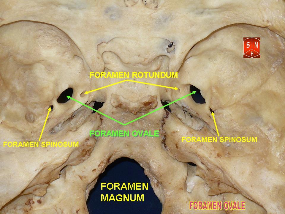
The foramen ovale (En: oval window) is a hole in the posterior part of the sphenoid bone, posterolateral to the foramen rotundum. It is one of the larger of the several holes (the foramina) in the skull. It transmits the mandibular nerve, a branch of the trigeminal nerve.
Structure
The foramen ovale is an opening in the greater wing of the sphenoid bone. The foramen ovale is one of two cranial foramina in the greater wing, the other being the foramen spinosum. The foramen ovale is posterolateral to the foramen rotundum and anteromedial to the foramen spinosum. Posterior and medial to the foramen is the opening for the carotid canal.
Contents
The following structures pass through foramen ovale:
- mandibular nerve (CN V) (a branch of the trigeminal nerve (CN V))
- accessory meningeal artery
- lesser petrosal nerve (a branch of the glossopharyngeal nerve)
- an emissary vein connecting the cavernous sinus with the pterygoid plexus
- (occasionally) meningeal branch of the mandibular nerve
Variation
In a study conducted on 100 skulls, the foramen ovale was divided into 2 or 3 components in 4.5% of the cases. The borders of the foramen in some skulls were also irregular and rough. This may suggest, based on radiological images, the presence of morbid changes, which might be the sole anatomical variation in the foramina ovalia of humans.
In newborn, the foramen ovale is about 3.85 mm and in the adults about 7.2 mm in length. The average maximal length is about 7.48 mm and its average minimal length is 4.17 mm in the adult. The width extends from 1.81 mm in the newborn to 3.7 mm in adults.
Development
Similar to other foramina, the foramen ovale differs in shape and size throughout life. In a study using over 350 skulls, the earliest perfect ring-shaped formation of the foramen ovale was observed in the 7th month of fetal life, and the latest in 3 years after birth.
Clinical significance
The foramen ovale is used as the entry point into the skull when conducting a percutaneous rhizotomy using either radio-frequency ablation, balloon compression or glycerol injection. These are performed to treat trigeminal neuralgia. In the procedure, the electrode is introduced through the cheek of an anesthetized patient and radiologically guided into the foramen ovale, with the intention of partially or fully ablating one or more of the divisions (typically the mandibular) to relieve pain.
This entry point is also used to surgically place local electrodes directly on the surface of the mesial temporal lobe, in order to observe neural activity of patients with suspected focal epilepsy.
History
Etymology
The name "foramen ovale" comes from the Latin "oval hole / window".
References
References
- Ray, Biswabina. (2005). "Anatomic variations of foramen ovale". Kathmandu University Medical Journal.
- Drake, Richard L.. (2005). "Gray's anatomy for students". Elsevier/Churchill Livingstone.
- Sinnatamby, Chummy S.. (2011). "Last's Anatomy".
- (2005). "The morphology and morphometry of the foramina of the greater wing of the human sphenoid bone". Folia Morphologica.
- Yanagi S. (1987). "Developmental studies on the foramen rotundum, foramen ovale and foramen spinosum of the human sphenoid bone". The Hokkaido Journal of Medical Science.
- (1984). "Postnatal enlargement of the foramina rotundum, ovale et spinosum and their topographical changes". Anatomischer Anzeiger.
- "Percutaneous stereotactic rhizotomy (PSR) for facial pain".
- (2006). "Foramen ovale electrodes can identify a focal seizure onset when surface EEG fails in mesial temporal lobe epilepsy". Epilepsia.
This article was imported from Wikipedia and is available under the Creative Commons Attribution-ShareAlike 4.0 License. Content has been adapted to SurfDoc format. Original contributors can be found on the article history page.
Ask Mako anything about Foramen ovale (skull) — get instant answers, deeper analysis, and related topics.
Research with MakoFree with your Surf account
Create a free account to save articles, ask Mako questions, and organize your research.
Sign up freeThis content may have been generated or modified by AI. CloudSurf Software LLC is not responsible for the accuracy, completeness, or reliability of AI-generated content. Always verify important information from primary sources.
Report