From Surf Wiki (app.surf) — the open knowledge base
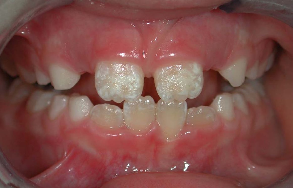
Enamel hypoplasia
Lack of tooth enamel
Lack of tooth enamel
| Field | Value |
|---|---|
| name | Enamel hypoplasia |
| image | 법랑질저형성증.jpg |
| field | Dentistry |
Enamel hypoplasia is a defect of the teeth in which the enamel is deficient in quantity, caused by defective enamel matrix formation during enamel development, as a result of inherited and acquired systemic condition(s). It can be identified as missing tooth structure and may manifest as pits or grooves in the crown of the affected teeth, and in extreme cases, some portions of the crown of the tooth may have no enamel, exposing the dentin. It may be generalized across the dentition or localized to a few teeth. Defects are categorized by shape or location. Common categories are pit-form, plane-form, linear-form, and localised enamel hypoplasia. Hypoplastic lesions are found in areas of the teeth where the enamel was being actively formed during a systemic or local disturbance. Since the formation of enamel extends over a long period of time, defects may be confined to one well-defined area of the affected teeth. Knowledge of chronological development of deciduous and permanent teeth makes it possible to determine the approximate time at which the developmental disturbance occurred. Enamel hypoplasia varies substantially among populations and can be used to infer health and behavioural impacts from the past. Defects have also been found in a variety of non-human animals.
Causes
Enamel hypoplasia is believed to result from the dysfunction of ameloblasts—enamel-producing cells—either for a short period of time or throughout their lifespan. Enamel hypoplasia has a wide variety of known causes. Some causes are hereditary and others are from environmental exposure. The severity and localization of disease presentation is dependent on the timing and stage of tooth development in which the defective enamel formation occurred.
Hereditary causes of enamel hypoplasia include:
- Primary abnormalities in enamel development, such as amelogenesis imperfecta
- Certain dermatological conditions, such as Ehlers-Danlos syndrome
- Other complex hereditary conditions such as Usher syndrome, Seckel syndrome, Treacher-Collins syndrome, Heimler syndrome & Ellis-Van Creveld syndrome, Down syndrome, Hypophosphatasia, Regional odontodysplasia, Cleft lip and palate, Vitamin D-resistant rickets.
Environmental causes of enamel hypoplasia include:
- Nutritional deficits, such as Vitamin D, iron or calcium deficiency
- Birth injury or preterm birth
- Smoking during pregnancy
- Trauma
- Radiation
- Certain infections (e.g. congenital syphilis, cytomegalovirus, congenital rubella)
- Certain diseases (such as undiagnosed and untreated celiac disease, chicken pox, congenital syphilis)
- High fever
- Hypoxia (i.e. from severe cardiac defect)
- Excessive fluoride exposure (dental fluorosis)
- Tetracycline
- Vitamin A, C, or D deficiency
Clinical relevance

Enamel hypoplasia is a risk factor for dental caries in children including early childhood caries (ECC), which continues to be a burden for many children. This association has been identified as significant and independent, and is believed that the formation of pits and missing enamel provides a suitable local environment for adhesion and colonization of cariogenic bacteria. Historically, enamel hypoplasia has been under-appreciated as a risk factor for ECC.
Developmental defects in enamel that affect tooth appearance in the esthetic zone (i.e. upper front teeth) may cause individuals to experience social embarrassment or anxiety regarding the appearance of their teeth.
Treatment
Dental sealants may be indicated for the prevention of caries in hypoplastic pit & fissure systems.
According to clinical trials, topical fluoride treatment in the form of gel or varnish is effective in preventing tooth decay in children with enamel defects. Treatment with other topical agents, such as calcium phosphate (CPP-ACP), may also be effective in the remineralization of areas with congenital or carious enamel defects.
In decayed teeth with enamel defects, only bonded restorations (e.g. glass ionomer, polyacid-modified composite resin and not amalgam) should be placed and cavity margins should not be placed in areas of defective enamel as this may lead to marginal leakage and recurrent decay. Stainless steel crowns with conservative preparation may be an effective method to eliminate sensitivity and prevent further breakdown in teeth with enamel defects requiring full-coverage crowns.
Other conditions affecting enamel development
Hypomineralization or hypocalcification, as opposed to hypoplasia, refers to a decrease in the mineral content of the enamel, not the total amount present. It can vary in its presentation, and teeth may appear visually normal or highly translucent. Affected enamel is softer and more susceptible to acid, wear and decay.
Molar-incisor hypomineralization
Molar-incisor hypomineralization (MIH) is a condition in which there are areas of hypomineralized or hypomatured enamel on the crowns of permanent molars and incisors. In these cases, teeth may lose their weakened enamel shortly after eruption and are highly susceptible to dental caries. Affected teeth are highly sensitive to chemical and thermal irritants, and may also be difficult to obtain anesthesia. The causes of MIH are thought to be similar to those of other forms of enamel hypoplasia, but occur concurrently with crown development in the permanent molars and incisors (birth to approximately 3 years of age).
Turner's hypoplasia
Turner's hypoplasia or Turner's tooth is a presentation of enamel hypoplasia that normally affects only a single tooth. Its causes can be the same as other forms of enamel hypoplasia, but it is most commonly associated with trauma to a primary maxillary central incisor and the subsequent developmental disturbance of the underlying permanent tooth. This may also result in dilaceration of the root of the permanent tooth.
References
References
- (January 2019). ["Dental anatomy, physiology, and occlusion"](https://archive.org/details/dentalanatomyphy00majo}}{{page needed). W.B. Saunders.
- (2018). "Severe Plane-Form Enamel Hypoplasia in a Dentition from Roman Britain". Dental Anthropology Journal.
- (April 2019). "A probable genetic origin for pitting enamel hypoplasia on the molars of Paranthropus robustus". Journal of Human Evolution.
- (September 1997). "Relationship of enamel hypoplasia to the pattern of tooth crown growth: a discussion". American Journal of Physical Anthropology.
- (2016). "An Enigmatic Hypoplastic Defect of the Maxillary Lateral Incisor in Recent and Fossil Orangutans from Sumatra (Pongo abelii) and Borneo (Pongo pygmaeus)". International Journal of Primatology.
- (2011). "Orban's Oral Histology and Embryology". Elsevier.
- (2000). "Interpreting Developmental Stress in Archaeological Pigs: The Chronology of Linear Enamel Hypoplasia". Journal of Archaeological Science.
- (April 2018). "Amelogenesis imperfecta in the dentition of a wild chimpanzee". Journal of Medical Primatology.
- (1991). "Occurrence of enamel hypoplasia in the dentitions of simian primates". Folia Primatologica; International Journal of Primatology.
- "Diagnosis of Celiac Disease". National Institute of Health (NIH).
- [http://celiac.nih.gov/PDF/Dental_Enamel_Defects_508.pdf Dental Enamel Defects and Celiac Disease] {{webarchive. link. (March 5, 2016 National Institute of Health (NIH))
- (March 2008). "Oral manifestations of celiac disease". Journal of Clinical Gastroenterology.
- bhong, akash. (2023-05-10). "Is Tooth decay hereditary? - Healthy bano".
- (2015). "Enamel hypoplasia and its role in identification of individuals: A review of literature". Indian Journal of Dentistry.
- [http://celiac.nih.gov/PDF/Dental_Enamel_Defects_508.pdf Dental Enamel Defects and Celiac Disease] {{Webarchive. link. (2016-03-05 National Institute of Health (NIH))
- (2012). "The oral manifestations of celiac disease: information for the pediatric dentist". Pediatric Dentistry.
- (2010). "Oral signs in the diagnosis of celiac disease: review of the literature". Minerva Stomatologica.
- [https://doctorstaci.com/enamel-hypoplasia/ Enamel Hypoplasia] {{Webarchive. link. (2021-06-07 Doctor Staci Whitman (2021))
- (July 2017). "Dental Enamel formation and Implicateions for Oral Health and Disease". Physiological Reviews.
- Seow, W. K.. (2014). "Developmental defects of enamel and dentine: challenges for basic science research and clinical management". Australian Dental Journal.
- (2008). "Esthetic and endosurgical management of Turner's hypoplasia; a sequlae of trauma to developing tooth germ". Journal of Indian Society of Pedodontics and Preventive Dentistry.
- (April 2009). "Hypoplasia of a Permanent Incisor Produced by Primary Incisor Intrusion: A Case Report". JCDA.
This article was imported from Wikipedia and is available under the Creative Commons Attribution-ShareAlike 4.0 License. Content has been adapted to SurfDoc format. Original contributors can be found on the article history page.
Ask Mako anything about Enamel hypoplasia — get instant answers, deeper analysis, and related topics.
Research with MakoFree with your Surf account
Create a free account to save articles, ask Mako questions, and organize your research.
Sign up freeThis content may have been generated or modified by AI. CloudSurf Software LLC is not responsible for the accuracy, completeness, or reliability of AI-generated content. Always verify important information from primary sources.
Report