From Surf Wiki (app.surf) — the open knowledge base
Contrast CT
Medical imaging technique

Medical imaging technique
Contrast CT, or contrast-enhanced computed tomography (CECT), is X-ray computed tomography (CT) using radiocontrast. Radiocontrasts for X-ray CT are generally iodine-based types. This is useful to highlight structures such as blood vessels that otherwise would be difficult to delineate from their surroundings. Using contrast material can also help to obtain functional information about tissues. Often, images are taken both with and without radiocontrast. CT images are called precontrast or native-phase images before any radiocontrast has been administered, and postcontrast after radiocontrast administration.
Bolus tracking

Bolus tracking is a technique to optimize timing of the imaging. A small bolus of radio-opaque contrast media is injected into a patient via a peripheral intravenous cannula. Depending on the vessel being imaged, the volume of contrast is tracked using a region of interest (abbreviated "R.O.I.") at a certain level and then followed by the CT scanner once it reaches this level. Images are acquired at a rate as fast as the contrast moving through the blood vessels.
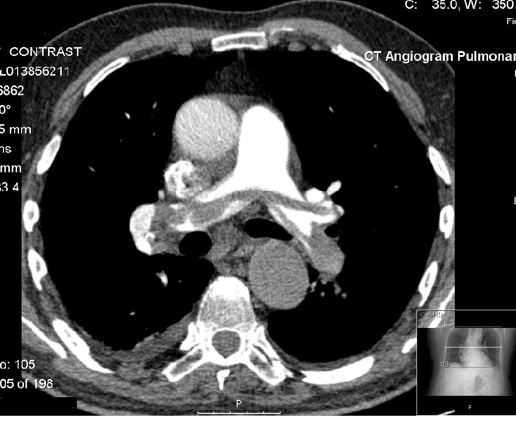
This method of imaging is used primarily to produce images of arteries, such as the aorta, pulmonary artery, cerebral, carotid and hepatic arteries.
Washout
"Washout" is where tissue loads radiocontrast during arterial phase, but then returns to a rather hypodense state in venous or later phases. This is a property of for example hepatocellular carcinoma as compared to the rest of the liver parenchyma.
Phases
Depending on the purpose of the investigation, there are standardized protocols for time intervals between intravenous radiocontrast administration and image acquisition, in order to visualize the dynamics of contrast enhancements in different organs and tissues. The main phases thereof are as follows:
| Phase | Time from injection | Time from bolus tracking | Targeted structures and findings | Non-enhanced CT (NECT) | Pulmonary arterial phase | Pulmonary venous phase | Early systemic arterial phase | Late systemicarterial phase | |||||||||||||
|---|---|---|---|---|---|---|---|---|---|---|---|---|---|---|---|---|---|---|---|---|---|
| Sometimes also called "arterial phase" or "early venous portal phase" | Pancreatic phase | Hepatic (most accurate) or late portal phase | Nephrogenic phase | Systemic venous phase | Delayed phase | ||||||||||||||||
| Sometimes called "wash out phase" or "equilibrium phase" | |||||||||||||||||||||
| title=5th European Conference of the International Federation for Medical and Biological Engineering 14 - 18 September 2011, Budapest, Hungary. Volume 37 of IFMBE Proceedings | author=Ákos Jobbágy | publisher=Springer Science & Business Media | year=2012 | isbn=978-3-642-23508-5}} | - | ||||||||||||||||
| 17–24 sec | - | ||||||||||||||||||||
| 15–20 sec | immediately | ||||||||||||||||||||
| 35–40 sec | 15–20 sec | ||||||||||||||||||||
| vauthors=Raman SP, Fishman EK | title=Advances in CT Imaging of GI Malignancies. | journal=Gastrointest Cancer Res | year= 2012 | volume= 5 | issue= 3 Suppl 1 | pages= S4-9 | pmid=22876336 | pmc=3413036 }} or 40 – 50 sec | 20–30 sec | ||||||||||||
| 70–80 sec | 50–60 sec | ||||||||||||||||||||
| 100 sec | 80 sec | ||||||||||||||||||||
| 180 sec | 160 sec | ||||||||||||||||||||
| date=December 2019 | reason=removed citation to predatory publisher content}} minutes | date=December 2019 | reason=removed citation to predatory publisher content}} minutes |
Angiography
Main article: CT angiography
CT angiography is a contrast CT taken at the location and corresponding phase of the blood vessels of interest, in order to detect vascular diseases. For example, an abdominal aortic angiography is taken in the arterial phase in the abdominal level, and is useful to detect for example aortic dissection.
Amount

Adults
The following table shows the preferable volume in normal weight adults. However, dosages may need to be adjusted or even withheld in patients with risks of iodinated contrast, such as hypersensitivity reactions, contrast-induced nephropathy, effects on thyroid function or adverse drug interactions.
| Exam | Iodine concentration | Comments | 300 mg/ml | 350 mg/ml | 370 mg/ml | |||
|---|---|---|---|---|---|---|---|---|
| CT of brain | url=http://www.medsafe.govt.nz/profs/Datasheet/o/Omnipaqueinj.pdf | title=New Zealand Datasheet | website=New Zealand Medicines and Medical Devices Safety Authority | access-date=2018-10-16}} | 80 ml | 75 ml | ||
| CT of thorax | Overall | 70–95 ml0.3–0.4 gI/kg in a 70kg individual, according to: | ||||||
| CT pulmonary angiogram | 20 mlUsing dual energy CTA (such as 90/150SnkVp), according to: | |||||||
| CT of abdomen | Overall | 70 ml | 60 ml | 55 ml | ||||
| Liver | 55 mlThe liver generally needs an enhancement of at least 30 HU for proper evaluation according to: | |||||||
| CT angiography | 25 mlCT-angiography in a 70kg person, with 100-150 mg I/kg by using 80 kVp, mAs-compensation for constant CNR, fixed injection duration adapted to scan time, automatic bolus tracking and a saline chaser, according to: |
The dose should be adjusted in those not having normal body weight, and in such cases the adjustment should be proportional to the lean body mass of the person. In obese patients, the Boer formula is the method of choice (at least in those with body mass index (BMI) between 35 and 40):
For men: Lean body mass = (0.407 × W) + (0.267 × H) − 19.2
For women: Lean body mass = (0.252 × W) + (0.473 × H) − 48.3
Children
Standard doses in children:
| Exam | Concentration of iodine | 300 mg/ml | 350 mg/ml |
|---|---|---|---|
| Generally | 2.0 ml/kg | 1.7 ml/kg | |
| CT of brain, neck or thorax | 1.5 ml/kg | 1.3 ml/kg |
Adverse effects
Iodinated contrast agents may cause allergic reactions, contrast-induced nephropathy, hyperthyroidism and possibly metformin accumulation. However, there are no absolute contraindications to iodinated contrast, so the benefits needs to be weighted against the risks.
As with CT scans in general, the radiation dose can potentially increase the risk of radiation-induced cancer.
The injection of iodinated contrast agents may sometimes lead to its extravasation.
Notes
References
References
- (2014). "Fundamentals of Body CT". Elsevier Health Sciences.
- (2000). "Detection and Characterisation of Renal Lesions by Multiphasic Helical Ct". Acta Radiologica.
- (2014). "CT and MR Imaging Diagnosis and Staging of Hepatocellular Carcinoma: Part II. Extracellular Agents, Hepatobiliary Agents, and Ancillary Imaging Features". Radiology.
- (2010). "Intravenous Contrast Medium Administration and Scan Timing at CT: Considerations and Approaches". Radiology.
- Robin Smithuis. "CT contrast injection and protocols".
- Ákos Jobbágy. (2012). "5th European Conference of the International Federation for Medical and Biological Engineering 14 - 18 September 2011, Budapest, Hungary. Volume 37 of IFMBE Proceedings". Springer Science & Business Media.
- Pavan Nandra. (2018). "Introducing the use of Flash CTPA; how does it compare to standard CTPA?".
- (2012). "Advances in CT Imaging of GI Malignancies.". Gastrointest Cancer Res.
- Otto van Delden and Robin Smithuis. "Pancreas - Carcinoma".
- Stuart E. Mirvis, Jorge A. Soto, Kathirkamanathan Shanmuganathan, Joseph Yu, Wayne S. Kubal. (2014). "Problem Solving in Emergency Radiology E-Book". Elsevier Health Sciences.
- "New Zealand Datasheet".
- (2018). "Lean Body Weight-Tailored Iodinated Contrast Injection in Obese Patient: Boer versus James Formula". BioMed Research International.
- (2010). "Multidetector CT in children: current concepts and dose reduction strategies". Pediatric Radiology.
- Stacy Goergen. "Iodine-containing contrast medium".
- (2019). "What you should know about prophylaxis and treatment of radiographic and magnetic resonance contrast medium extravasation". Acta Radiol.
This article was imported from Wikipedia and is available under the Creative Commons Attribution-ShareAlike 4.0 License. Content has been adapted to SurfDoc format. Original contributors can be found on the article history page.
Ask Mako anything about Contrast CT — get instant answers, deeper analysis, and related topics.
Research with MakoFree with your Surf account
Create a free account to save articles, ask Mako questions, and organize your research.
Sign up freeThis content may have been generated or modified by AI. CloudSurf Software LLC is not responsible for the accuracy, completeness, or reliability of AI-generated content. Always verify important information from primary sources.
Report