From Surf Wiki (app.surf) — the open knowledge base
Axillary artery
Large blood vessel bringing oxygenated blood to the thorax
Large blood vessel bringing oxygenated blood to the thorax
| Field | Value |
|---|---|
| Name | Axillary artery |
| Latin | arteria axillaris |
| Image | Axillary limits.PNG |
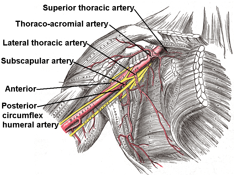
| Caption | Axillary artery and its branches—anterior view of right upper limb and thorax. Upper and lower limits labeled. |
| Image2 | Pectoralis minor.svg |
| Caption2 | The pectoralis minor muscle is used as a landmark for dividing the axillary artery into three parts. |
| BranchFrom | Subclavian artery |
| BranchTo | Superior thoracic |
| thoracoacromial | |
| lateral thoracic | |
| subscapular | |
| anterior circumflex humeral | |
| posterior circumflex humeral | |
| continues as brachial artery | |
| Vein | Axillary vein |
| Supplies | Axilla |
thoracoacromial lateral thoracic subscapular anterior circumflex humeral posterior circumflex humeral continues as brachial artery
In human anatomy, the axillary artery is a large blood vessel that conveys oxygenated blood to the lateral aspect of the thorax, the axilla (armpit) and the upper limb. Its origin is at the lateral margin of the first rib, before which it is called the subclavian artery.
After passing the lower margin of teres major it becomes the brachial artery.
Structure
The axillary artery is often referred to as having three parts, with these divisions based on its location relative to the pectoralis minor muscle, which is superficial to the artery.
- First part – the part of the artery superior to the pectoralis minor
- Second part – the part of the artery posterior to the pectoralis minor
- Third part – the part of the artery inferior to the pectoralis minor.
Relations
The axillary artery is accompanied by the axillary vein, which lies medial to the artery, along its length.
In the axilla, the axillary artery is surrounded by the brachial plexus. The second part of the axillary artery is the reference for the locational descriptions of the cords in the brachial plexus. For example, the posterior cord of the brachial plexus is so named because it lies posterior to the second part of the artery.
Branches
The axillary artery has several smaller branches. The branches can be remembered, in order, when traveling from the heart, with the mnemonics "Seek The Lord, Serve All People", "Summertime: The Lakers Schedule Another Parade", "Some Traumas Leave Scars And Pain", or "She Tastes Like Sweet Apple Pie." The origin of these branches is highly variable (e.g. the posterior and anterior circumflex arteries often have a common trunk). An arterial branch is named for its course, not its origin.
- First part (1 branch)
- Superior thoracic artery (Supreme thoracic artery)
- Second part (2 branches)
- Thoraco-acromial artery
- Lateral thoracic artery. If the lateral thoracic artery is not branching from the axillary artery, will most likely branch from the following (in order of likelihood): (1) thoracoacromial, (2) third part of axillary artery, (3) suprascapular artery, (4) subscapular artery
- Third part (3 branches)
- Subscapular artery
- Anterior humeral circumflex artery
- Posterior humeral circumflex artery
Continues as the brachial artery past the inferior border of the teres major.
Clinical significance
The axillary artery can be safely clamped without endangering the arm, but only in a location proximal to the origin of the subscapular artery (and distal to the thyrocervical trunk of the subclavian artery). The anastomotic network surrounding the scapula provides an alternate path for collateral circulation to the arm from arteries including the dorsal scapular artery and suprascapular artery.
The right axillary artery is often used as an arterial cannulation site in cardiac surgery, particularly for repair of aortic dissection and replacement of the ascending aorta and aortic arch.
Additional images
Image:Gray576.png|The veins of the right axilla, viewed from in front. Image:Gray809.png|The right brachial plexus (infraclavicular portion) in the axillary fossa; viewed from below and in front. Image:Gray810.png|Suprascapular and axillary nerves of right side, seen from behind. File:PLEXUS BRACHIALIS.jpg|Brachial plexus and axillary artery File:Slide3v.JPG|Axillary artery File:Slide2bbbb.JPG|Axillary artery File:Slide9JJJ.JPG|Axillary artery File:Slide11OOO.JPG|Axillary artery File:Slide15SSS.JPG|Axillary artery File:Slide5EEEE.JPG|Axillary artery
References
References
- (February 2022). "The "Hand as Foot" teaching method in axillary artery anatomy". Asian Journal of Surgery.
- (2011). "Anatomy for diagnostic imaging". Elsevier Ltd.
- {{MedicalMnemonics. 1208. 852. 663
This article was imported from Wikipedia and is available under the Creative Commons Attribution-ShareAlike 4.0 License. Content has been adapted to SurfDoc format. Original contributors can be found on the article history page.
Ask Mako anything about Axillary artery — get instant answers, deeper analysis, and related topics.
Research with MakoFree with your Surf account
Create a free account to save articles, ask Mako questions, and organize your research.
Sign up freeThis content may have been generated or modified by AI. CloudSurf Software LLC is not responsible for the accuracy, completeness, or reliability of AI-generated content. Always verify important information from primary sources.
Report