From Surf Wiki (app.surf) — the open knowledge base
Atypical ductal hyperplasia
| Field | Value |
|---|---|
| name | Atypical ductal hyperplasia |
| image | Atypical ductal hyperplasia - very low mag.jpg |
| width | 250 |
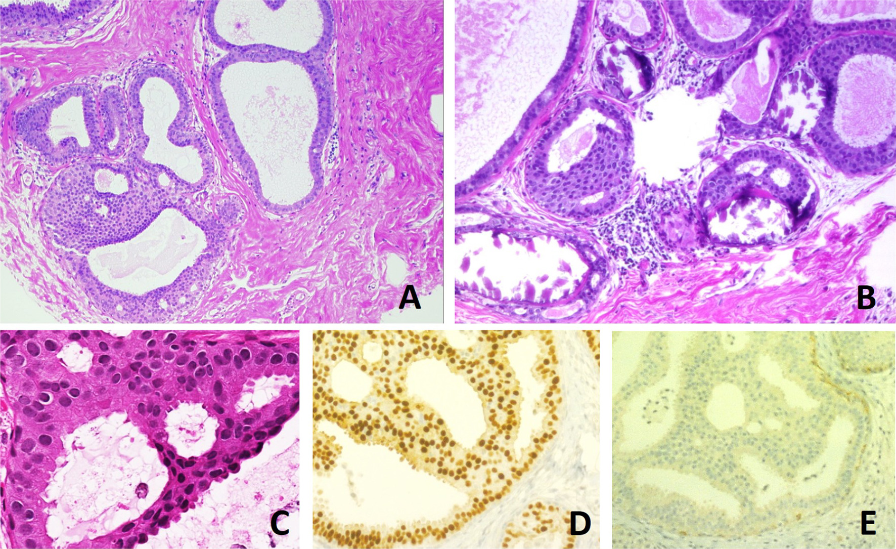
| caption | Very low magnification micrograph of atypical ductal hyperplasia (ADH). The piece with ADH was circled by the pathologist with a marker, as it is so small, and sent for an additional opinion. H&E stain. |
| field | Gynecology, pathology |
Atypical ductal hyperplasia (ADH) is the term used for a benign lesion of the breast that indicates an increased risk of breast cancer.
The name of the entity is descriptive of the lesion; ADH is characterized by cellular proliferation (hyperplasia) within one or two breast ducts and (histomorphologic) architectural abnormalities, i.e. the cells are arranged in an abnormal or atypical way, more so than usual ductal hyperplasia.
In the context of a core (needle) biopsy, ADH is considered an indication for a breast lumpectomy, also known as a surgical (excisional) biopsy, to exclude the presence of breast cancer.
Signs and symptoms
ADH, generally, is asymptomatic. It usually comes to medical attention on a screening mammogram, as a non-specific suspicious abnormality that requires a biopsy.
Pathology
ADH, cytologically, architecturally and on a molecular basis, is identical to a low-grade ductal carcinoma in situ (DCIS); however, it has a limited extent, i.e. is present in a very small amount ( Image:Atypical ductal hyperplasia - low mag.jpg | Low mag. Image:Atypical ductal hyperplasia - high mag.jpg | High mag.
Relation to low-grade ductal carcinoma in situ
While the histopathologic features and molecular features of ADH are that of (low-grade) DCIS, its clinical behaviour, unlike low-grade DCIS, is substantially better; thus, the more aggressive treatment for DCIS is not justified.
Diagnosis
It is diagnosed based on tissue, e.g. a biopsy, showing ductal hyperplasia.
There is no single definite cutoff that separates atypical ductal hyperplasia from ductal carcinoma in situ, but the following are important distinctive features of atypical ductal hyperplasia, with suggested cutoffs:
- Size less than 2 mm.
- Not involving more than one duct.
- The atypical epithelial proliferation is admixed with a second population of proliferative cells without atypia.
- The proliferation completely involves the terminal ductal lobular unit(s), to a limited extent.
Treatment
ADH, if found on a surgical (excisional) biopsy of a mammographic abnormality, does not require any further treatment, only mammographic follow-up.
If ADH is found on a core (needle) biopsy (a procedure which generally does not excise a suspicious mammographic abnormality), a surgical biopsy, i.e. a breast lumpectomy, to completely excise the abnormality and exclude breast cancer is the typical recommendation.
Prognosis
Cancer risk for ADH on a core biopsy
The rate at which breast cancer (ductal carcinoma in situ or invasive mammary carcinoma (all breast cancer except DCIS and LCIS)) is found at the time of a surgical (excisional) biopsy, following the diagnosis of ADH on a core (needle) biopsy varies considerably from hospital-to-hospital (range 4-54%). In two large studies, the conversion of an ADH on core biopsy to breast cancer on surgical excision, known as "up-grading", is approximately 30%.
Cancer risk based on follow-up
The relative risk of breast cancer based on a median follow-up of 8 years, in a case control study of US registered nurses, is 3.7.
References
References
- "Understanding Breast Changes - National Cancer Institute".
- (May 1995). "Atypical ductal hyperplasia diagnosed at stereotaxic core biopsy of breast lesions: an indication for surgical biopsy". AJR Am J Roentgenol.
- (Dec 2006). "Discrepancies in the diagnosis of intraductal proliferative lesions of the breast and its management implications: results of a multinational survey". Virchows Arch.
- (2018). "Atypical ductal hyperplasia and the risk of underestimation: tissue sampling method, multifocality, and associated calcification significantly influence the diagnostic upgrade rate based on subsequent surgical specimens". Breast Cancer.
- (January 2009). "Frequency and upgrade rates of atypical ductal hyperplasia diagnosed at stereotactic vacuum-assisted breast biopsy: 9-versus 11-gauge". AJR Am J Roentgenol.
- (2016). "Atypical Ductal Hyperplasia Bordering on Ductal Carcinoma In Situ". International Journal of Surgical Pathology.
- (Feb 2011). "Factors associated with upgrading to malignancy at surgery of atypical ductal hyperplasia diagnosed on core biopsy". Breast.
- (Oct 2006). "Correlation between core biopsy and excisional biopsy in breast high-risk lesions". Am J Surg.
- (Feb 1992). "A prospective study of benign breast disease and the risk of breast cancer". JAMA.
This article was imported from Wikipedia and is available under the Creative Commons Attribution-ShareAlike 4.0 License. Content has been adapted to SurfDoc format. Original contributors can be found on the article history page.
Ask Mako anything about Atypical ductal hyperplasia — get instant answers, deeper analysis, and related topics.
Research with MakoFree with your Surf account
Create a free account to save articles, ask Mako questions, and organize your research.
Sign up freeThis content may have been generated or modified by AI. CloudSurf Software LLC is not responsible for the accuracy, completeness, or reliability of AI-generated content. Always verify important information from primary sources.
Report