From Surf Wiki (app.surf) — the open knowledge base
Aortic arch
Part of the aorta
Part of the aorta
| Field | Value |
|---|---|
| Name | Aortic arch |
| Latin | arcus aortae |
| Image | Gray506.svg |
| Caption | The aortic arch has three branches, the brachiocephalic trunk, left common carotid artery, and left subclavian artery. |
| Image2 | Gray505.png |
| Caption2 | The aortic arch and its branches shown in situ. |
| BranchFrom | Ascending aorta |
| Vein | Combination of superior and inferior vena cava |
| Precursor | Fourth left pharyngeal arch artery |
| Supplies | From its branches, the upper body, arms, head and neck. As a part of the aorta, the entire body, with exception of the respiratory zone of the lung and the heart. |
- Brachiocephalic trunk
- Left common carotid artery
- Left subclavian artery Continues as descending aorta, thoracic part
The aortic arch, arch of the aorta, or transverse aortic arch () is the part of the aorta between the ascending and descending aorta. The arch travels backward, so that it ultimately runs to the left of the trachea.
Structure
The aorta begins at the level of the upper border of the second/third sternocostal articulation of the right side, behind the ventricular outflow tract and pulmonary trunk. The right atrial appendage overlaps it. The first few centimeters of the ascending aorta and pulmonary trunk lies in the same pericardial sheath and runs at first upward, arches over the pulmonary trunk, right pulmonary artery, and right main bronchus to lie behind the right second coastal cartilage. The right lung and sternum lies anterior to the aorta at this point. The aorta then passes posteriorly and to the left, anterior to the trachea, and arches over left main bronchus and left pulmonary artery, and reaches to the left side of the T4 vertebral body. Apart from T4 vertebral body, other structures such as trachea, oesophagus, and thoracic duct (from front to back) also lies to the left of the aorta. Inferiorly, the arch of aorta is connected to ligamentum arteriosum while superiorly, it gives rise to three main branches. The arch of aorta continues as the descending aorta after T4 vertebral body.
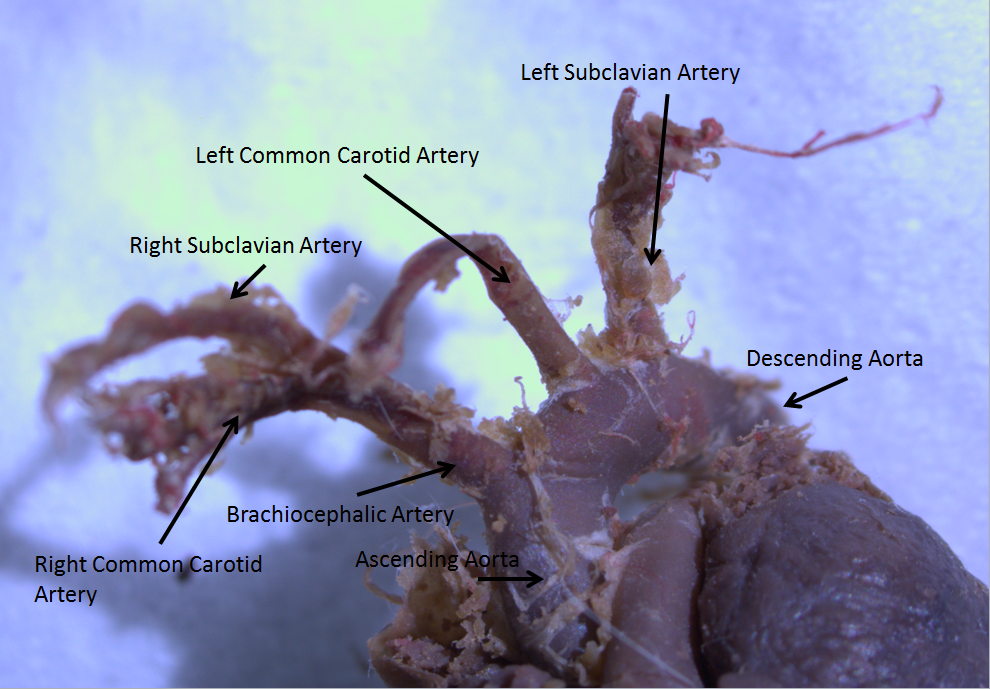
The aortic arch has three main branches on its superior aspect. The first, and largest, branch of the arch of the aorta is the brachiocephalic trunk, which is to the right and slightly anterior to the other two branches and originates behind the manubrium of the sternum. Next, the left common carotid artery originates from the aortic arch to the left of the brachiocephalic trunk, then ascends along the left side of the trachea and through the superior mediastinum. Finally, the left subclavian artery comes off of the aortic arch to the left of the left common carotid artery and ascends, with the left common carotid, through the superior mediastinum and along the left side of the trachea. An anatomical variation is that the left vertebral artery can arise from the aortic arch instead of the left subclavian artery.
The arch of the aorta forms two curvatures: one with its convexity upward, the other with its convexity forward and to the left. Its upper border is usually about 2.5 cm below the superior border to the manubrium sterni. Blood flows from the upper curvature to the upper regions of the body, located above the heart - namely the arms, neck, and head.
Coming out of the heart, the thoracic aorta has a maximum diameter of 40 mm at the root. By the time it becomes the ascending aorta, the diameter should be
The arch of the aorta lies within the mediastinum.
At the cellular level, the aorta and the aortic arch are composed of three layers: The tunica intima, which surrounds the lumen and is composed of simple squamal epithelial cells; the tunica media, composed of smooth cell muscles and elastic fibers; and, the tunica adventitia, composed of loose collagen fibers. Innervated by barometric nerve terminals, the aortic arch is responsible for sensing changes in the dilation of the vascular walls, inducing changes in heart rate to compensate for changes in blood pressure.
Development
The aortic arch is the connection between the ascending and descending aorta, and its central part is formed by the left 4th aortic arch during early development.
The ductus arteriosus connects to the lower part of the arch in foetal life. This allows blood from the right ventricle to mostly bypass the pulmonary vessels as they develop.
The final section of the aortic arch is known as the aortic isthmus. This is so called because it is a narrowing (isthmus) of the aorta as a result of decreased blood flow when in foetal life. As the left ventricle of the heart increases in size throughout life, the narrowing eventually dilates to become a normal size. If this does not occur, this can result in coarctation of the aorta. The ductus arteriosus connects to the final section of the arch in foetal life. Ductus arteriosus then regresses to become ligamentum arteriosum during later life.
Variation
There are three common variations in how arteries branch from the aortic arch. In about 75% of individuals, the branching is "normal", as described above. In some individuals the left common carotid artery originates from the brachiocephalic artery rather than the aortic arch. In others, the brachiocephalic artery and left common carotid artery share an origin. This variant is found in approximately a 20% of the population. In a third variant, the brachiocephalic artery splits into three arteries: the left common carotid artery, the right common carotid artery and the right subclavian artery; this variant is found in an estimated 7% of individuals. In rare cases, the thyroid ima artery, a variant artery supplying the thyroid gland may arise from the aortic arch.
Clinical significance
The aortic knob is the prominent shadow of the aortic arch on a frontal chest radiograph.
Aortopexy is a surgical procedure in which the aortic arch is fixed to the sternum in order to keep the trachea open.
Aortic isthmus is the relatively fixed part of the aortic arch. It is prone to shearing force and trauma that can cause it to tear and result in massive bleeding.
Additional images
Image:Gray490.png|The arch of aorta can be seen here, with the lungs to either side and emerging from the heart, below. Image:Gray793.png|A branch of the vagus nerve, the recurrent laryngeal nerve, passes underneath the arch of aorta. The nerve is seen here.
References
References
- ''[[OED]]'' 2nd edition, 1989, as {{IPA. /eɪ'ɔ:ɹtɪk/.
- [https://www.merriam-webster.com/dictionary/aortic Entry "aortic"] in ''[https://www.merriam-webster.com/ Merriam-Webster Online Dictionary]''.
- (2011). "Anatomy for diagnostic imaging". Elsevier Ltd.
- S. Standring. "Gray's Anatomy The Anatomical Basis Of Clinical Practice, 40th Edition". Elsevier Health Sciences UK.
- (2011). "Textbook of anatomy". Jaypee Brothers Medical Publishers.
- Drake, Richard L.. (2005). "Gray's anatomy for students". Elsevier/Churchill Livingstone.
- (2006). "Clinical anatomy for students : problem solving approach". Jaypee Bros. Medical Publishers.
- (2008). "Normal thoracic aorta diameter on cardiac computed tomography in healthy asymptomatic adults: impact of age and gender". Acad Radiol.
- (2008). "Aortic size assessment by noncontrast cardiac computed tomography: normal limits by age, gender, and body surface area". JACC Cardiovasc Imaging.
- "The Cardiovascular System (Blood Vessels)".
- webmaster@studentconsult.com. "Printed from STUDENT CONSULT: Berne and Levy Physiology 6E - The Online Medical Library for Students plus USMLE Steps 123 (ver. 2.9)".
- (2013-03-01). "Clarification of the identity of the mammalian fifth pharyngeal arch artery". Clinical Anatomy.
- (2016). "The Aortic Isthmus: A Significant yet Underexplored Watershed of the Fetal Circulation". Fetal Diagnosis and Therapy.
- (2008). "Rubin's Pathology: clinicopathologic foundations of medicine". Wolters Kluwer/Lippincott Williams & Wilkins.
- (2007). "Computed tomography and magnetic resonance of the thorax". Wolters Kluwer/Lippincott Williams & Wilkins.
- (2012). "Letters to Editor Bovine arch". Archives of Medical Science.
- (1989). "[A case of the thyroidea ima artery arising from the aortic arch]". Kaibogaku Zasshi. Journal of Anatomy.
- [http://www.wrongdiagnosis.com/medical/aortic_knob.htm wrongdiagnosis.com > Aortic knob] Citing: Stedman's Medical Spellchecker, 2006 Lippincott Williams & Wilkins.
This article was imported from Wikipedia and is available under the Creative Commons Attribution-ShareAlike 4.0 License. Content has been adapted to SurfDoc format. Original contributors can be found on the article history page.
Ask Mako anything about Aortic arch — get instant answers, deeper analysis, and related topics.
Research with MakoFree with your Surf account
Create a free account to save articles, ask Mako questions, and organize your research.
Sign up freeThis content may have been generated or modified by AI. CloudSurf Software LLC is not responsible for the accuracy, completeness, or reliability of AI-generated content. Always verify important information from primary sources.
Report