From Surf Wiki (app.surf) — the open knowledge base
Actinomycosis
| Field | Value |
|---|---|
| image | Actinomycosis PHIL 2856 lores.jpg |
| caption | A man with actinomycosis on the right side of his face |
| field | Infectious disease |
|
Actinomycosis is a rare and chronic infectious bacterial disease caused by the gram-positive Actinomyces species. The name refers to ray-like appearance of the organisms in the granules. About 70% of infections are due to either Actinomyces israelii or A. gerencseriae.
Signs and symptoms
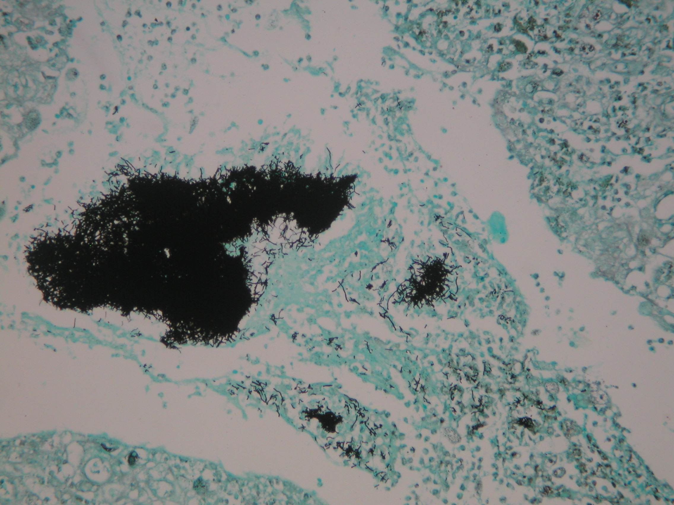
The disease is characterised by the formation of painful abscesses in the mouth, lungs, breast, or gastrointestinal tract. Actinomycosis abscesses grow larger as the disease progresses, often over months. In severe cases, they may penetrate the surrounding bone and muscle to the skin, where they break open and leak large amounts of pus, which often contains characteristic granules filled with progeny bacteria. These granules are often called "sulfur granules" due to their yellow appearance, although they may also be white, gray or brown.{{Cite book | author1-first = Robert M. | author1-last = Kliegman | author2-first = Joseph W., III | author2-last = St. Geme
Causes

Actinomycosis is primarily caused by any of several members of the bacterial genus Actinomyces. These bacteria are generally anaerobes. In animals, they normally live in the small spaces between the teeth and gums, causing infection only when they can multiply freely in anoxic environments. An affected human often has recently had dental work, poor oral hygiene, periodontal disease, radiation therapy, or trauma (broken jaw) causing local tissue damage to the oral mucosa, all of which predispose the person to developing actinomycosis. A. israelii is a normal commensal species part of the microbiota species of the lower reproductive tract of women. They are also normal commensals among the gut flora of the caecum; thus, abdominal actinomycosis can occur following removal of the appendix. The three most common sites of infection are decayed teeth, the lungs, and the intestines. Actinomycosis infections are typically polymicrobial, containing additional bacterial species; as Actinomyces itself has little invasive ability, these other species often aid in the infection process.{{Cite book | author1-last = Schaal | author1-first = Klaus P. | author2-last = Yassin | author2-first = Atteyet F. | author3-last = Stackebrandt | author3-first = E. R. K. O. | chapter-url = https://books.google.com/books?id=swciHNNWZDEC&pg=PA485
Actinomycosis (5287304877).jpg Actinomycosis (5287905500).jpg Actinomycosis - Gram stain (5285453121).jpg Actinomycosis - Gram stain (5286050280).jpg Actinomycosis - Gram stain (5286050326).jpg
Diagnosis
The diagnosis of actinomycosis can be a difficult one to make. In addition to microbiological examinations, magnetic resonance imaging and immunoassays may be helpful.
Treatment
Actinomyces bacteria are generally sensitive to penicillin, which is frequently used to treat actinomycosis. In cases of penicillin allergy, doxycycline is used. Sulfonamides such as sulfamethoxazole may be used as an alternative regimen at a total daily dosage of 2–4 grams. Response to therapy is slow and may take months. Hyperbaric oxygen therapy may also be used as an adjunct to conventional therapy when the disease process is refractory to antibiotics and surgical treatment.
Epidemiology
Disease incidence is greater in males between the ages of 20 and 60 years than in females. Before antibiotic treatments became available, the incidence in the Netherlands and Germany was one per 100,000 people/year. Incidence in the U.S. in the 1970s was one per 300,000 people/year, while in Germany in 1984, it was estimated to be one per 40,000 people/year. The use of intrauterine devices (IUDs) has increased incidence of genitourinary actinomycosis in females. -- Incidence of oral actinomycosis, which is harder to diagnose, has increased.
History
In 1877, pathologist Otto Bollinger described the presence of A. bovis in cattle, and shortly afterwards, James Israel discovered A. israelii in humans. In 1890, Eugen Bostroem isolated the causative organism from a culture of grain, grasses, and soil. After Bostroem's discovery, a general misconception existed that actinomycosis was a mycosis that affected individuals who chewed grass or straw. The pathogen is still known as the “great masquerader". Bergey's Manual of Systematic Bacteriology classified the organism as bacterial in 1939, but the disease remained classified as a fungus in the 1955 edition of the Control of Communicable Diseases in Man.
Violinist Joseph Joachim died of actinomycosis on 15 August 1907. The Norwegian painter Halfdan Egedius died from actinomycosis on 2 February 1899.
Other animals
Main article: Actinomycosis in animals
Actinomycosis occurs rarely in humans, but rather frequently in cattle as a disease called "lumpy jaw". This name refers to the large abscesses that grow on the head and neck of the infected animal. It can also rarely affect sheep, swine, horses, dogs, and other mammals.
References
References
- (2014). "Actinomycosis: etiology, clinical features, diagnosis, treatment, and management". Infect Drug Resist.
- Bowden GHW. (1996). "Actinomycosis ''in:'' Baron's Medical Microbiology". Univ of Texas Medical Branch.
- Brook, I. (Oct 2008). "Actinomycosis: diagnosis and management.". Southern Medical Journal.
- Mabeza, GF. (March 2003). "Pulmonary actinomycosis". European Respiratory Journal.
- (1 January 2015). "Primary actinomycosis of the breast caused by Actinomyces turicensis with associated Peptoniphilus harei". Breast Disease.
- (2004). "Sherris Medical Microbiology". McGraw Hill.
- Hoffman, Barbara. (2012). "Williams gynecology". McGraw-Hill Medical.
- Böhm, Ingrid. (October 2006). "Magnetic Resonance Imaging Meets Immunology: An Unusual Combination of Diagnostic Tools Leads to the Diagnosis Actinomycosis". The American Journal of Gastroenterology.
- "Bone Infections". US National Library of Medicine.
- "Osteomyelitis (Refractory)".
- (2007). "Fitzpatrick's Dermatology in General Medicine". McGraw Hill.
- (12 May 2010). "Bacteria That Masquerade as Fungi: Actinomycosis/Nocardia". Proceedings of the American Thoracic Society.
- (1944). "Stitt's Diagnosis, Prevention and Treatment of Tropical Diseases".
- (1955). "Control of Communicable Diseases in Man". American Public Health Association.
- Smith GW. (July 2020). "Actinomycosis in Cattle, Swine, and Other Animals". MSD Manual.
This article was imported from Wikipedia and is available under the Creative Commons Attribution-ShareAlike 4.0 License. Content has been adapted to SurfDoc format. Original contributors can be found on the article history page.
Ask Mako anything about Actinomycosis — get instant answers, deeper analysis, and related topics.
Research with MakoFree with your Surf account
Create a free account to save articles, ask Mako questions, and organize your research.
Sign up freeThis content may have been generated or modified by AI. CloudSurf Software LLC is not responsible for the accuracy, completeness, or reliability of AI-generated content. Always verify important information from primary sources.
Report