From Surf Wiki (app.surf) — the open knowledge base
Hemorheology
Study of flow properties of blood and its elements of plasma and cells
Study of flow properties of blood and its elements of plasma and cells
Hemorheology, also spelled haemorheology (haemo from Greek 'αἷμα, haima 'blood'; and rheology, from Greek ῥέω rhéō, 'flow' and -λoγία, -logia 'study of'), or blood rheology, is the study of flow properties of blood and its elements of plasma and cells. Proper tissue perfusion can occur only when blood's rheological properties are within certain levels. Alterations of these properties play significant roles in disease processes.{{cite book | url-access = limited
Blood viscosity
Blood viscosity is a measure of the resistance of blood to flow. It can also be described as the thickness and stickiness of blood. This biophysical property makes it a critical determinant of friction against the vessel walls, the rate of venous return, the work required for the heart to pump blood, and how much oxygen is transported to tissues and organs. These functions of the cardiovascular system are directly related to vascular resistance, preload, afterload, and perfusion, respectively.
The primary determinants of blood viscosity are hematocrit, red blood cell deformability, red blood cell aggregation, and plasma viscosity. Plasma's viscosity is determined by water-content and macromolecular components, so these factors that affect blood viscosity are the plasma protein concentration and types of proteins in the plasma.{{cite journal |archive-url=http://arquivo.pt/wayback/20160514165952/http://iospress.metapress.com/openurl.asp?genre=article&issn=1386-0291&volume=39&issue=1&spage=243 |url-status=dead |archive-date=2016-05-14
Clinical significance
Many conventional cardiovascular risk factors have been independently linked to whole blood viscosity.
| Cardiovascular risk factors linked independently to whole blood viscosity{{cite journal |
|---|
| s2cid=6366788 |
| Hypertension |
| Total cholesterol |
| VLDL-cholesterol |
| LDL-cholesterol |
| HDL-cholesterol (negative correlation) |
| Triglycerides |
| Chylomicrons |
| Diabetes mellitus and insulin resistance |
| Metabolic syndrome |
| Obesity |
| Cigarette smoking |
| Male sex |
| Age |
Anemia can reduce blood viscosity, which may lead to heart failure. Furthermore, elevation of plasma viscosity correlates to the progression of coronary and peripheral artery diseases.
Normal level
In pascal-seconds (Pa·s), the viscosity \mu of blood at 37 °C is normally 3 × 10⁻³ to 4 × 10⁻³, respectively 3 - 4 centipoise (cP) in the centimetre gram second system of units.
\mu = (3 \sim 4) \cdot 10^{-3} , Pa \cdot s
\nu = \frac{\mu}{\rho} = \frac{(3 \sim 4) \cdot 10^{-3} Pa\cdot s}{1.06\cdot 10^{3} \frac{kg}{m^3}} = (2.8 \sim 3.8) \cdot 10^{-6} , \frac{m^2}{s},
where \rho is the density. Blood viscosity can be measured by viscometers capable of measurements at various shear rates, such as a rotational viscometer.{{cite journal |hdl-access=free
Blood viscoelasticity
Blood is a viscoelastic fluid, meaning that it possesses both viscous and fluid characteristics. The viscous component arises primarily through the viscosity of blood plasma, while the elastic component arises from deformation of the red blood cells. As the heart contracts, mechanical energy is transferred from the heart to the blood; a small part of the energy is dissipated by the viscosity of the suspension, another part is stored as elastic energy in the red blood cells, and the remaining energy is used to drive blood circulation and is thus converted to kinetic energy. Viscoelastic fluids make up a larger class of fluids called non-Newtonian fluids.
The red blood cells occupy about half of the volume of blood and possess elastic properties. This elastic property is the largest contributing factor to the viscoelastic behavior of blood. The large volume percentage of red blood cells at a normal hematocrit level leaves little room for cell motion and deformation without interacting with a neighboring cell. Calculations have shown that the maximum volume percentage of red blood cells without deformation is 58% which is in the range of normally occurring levels.{{cite book
When the red cells are at rest or at very small shear rates, they tend to aggregate and stack together in an energetically favorable manner. The attraction is attributed to charged groups on the surface of cells and to the presence of fibrinogen and globulins. This aggregated configuration is an arrangement of cells with the least amount of deformation. With very low shear rates, the viscoelastic property of blood is dominated by the aggregation and cell deformability is relatively insignificant. As the shear rate increases the size of the aggregates begins to decrease. With a further increase in shear rate, the cells will rearrange and orient to provide channels for the plasma to pass through and for the cells to slide. In this low to medium shear rate range, the cells wiggle with respect to the neighboring cells allowing flow. The influence of aggregation properties on the viscoelasticity diminish and the influence of red cell deformability begins to increase. As shear rates become large, red blood cells will stretch or deform and align with the flow. Cell layers are formed, separated by plasma, and flow is now attributed to layers of cells sliding on layers of plasma. The cell layer allows for easier flow of blood and as such there is a reduced viscosity and reduced elasticity. The viscoelasticity of the blood is dominated by the deformability of the red blood cells.
Maxwell model
Maxwell Model concerns Maxwell fluids or Maxwell material. The material in Maxwell Model is a fluid which means it respects continuity properties for conservative equations : Fluids are a subset of the phases of matter and include liquids, gases, plasmas and, to some extent, plastic solids. Maxwell model is made to estimate local conservative values of viscoelasticity by a global measure in the integral volume of the model to be transposed to different flow situations. Blood is a complex material where different cells like red blood cells are discontinuous in plasma. Their size and shape are irregular too because they are not perfect spheres. Complicating moreover blood volume shape, red cells are not identically distributed in a blood sample volume because they migrate with velocity gradients in direction to the highest speed areas calling the famous representation of the Fåhræus–Lindqvist effect, aggregate or separate in sheath or plug flows described by Thurston. Typically, the Maxwell Model described below is uniformly considering the material (uniform blue color) as a perfect distributed particles fluid everywhere in the volume (in blue) but Thurston reveals that packs of red cells, plugs, are more present in the high speed region, if y is the height direction in the Maxwell model figure, (y~H) and there is a free cells layer in the lower speed area (y~0) what means the plasma fluid phase that deforms under Maxwell Model is strained following inner linings that completely escape from the analytical model by Maxwell.
In theory, a fluid in a Maxwell Model behaves exactly similarly in any other flow geometry like pipes, rotating cells or in rest state. But in practice, blood properties vary with the geometry and blood has shown being an inadequate material to be studied as a fluid in common sense. So Maxwell Model gives trends that have to be completed in real situation followed by Thurston model in a vessel regarding distribution of cells in sheath and plug flows.
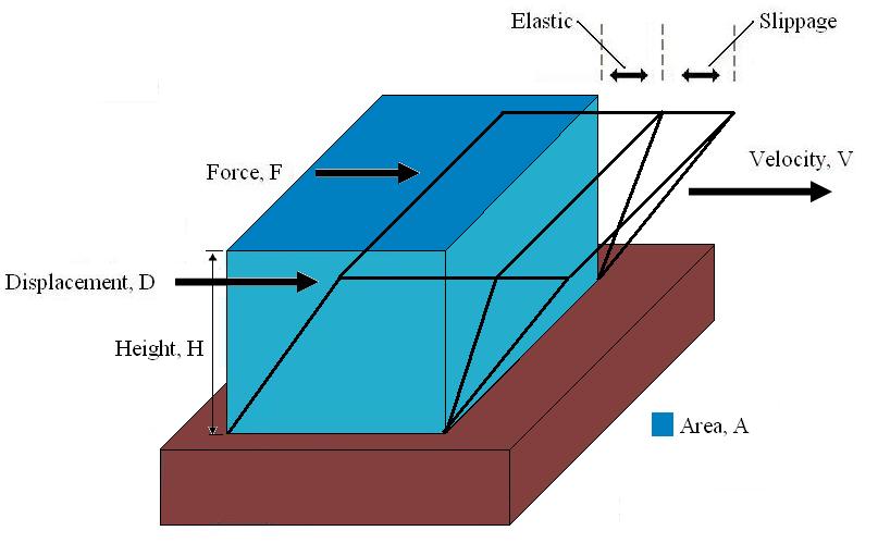
If a small cubical volume of blood is considered, with forces being acted upon it by the heart pumping and shear forces from boundaries. The change in shape of the cube will have 2 components:
- Elastic deformation which is recoverable and is stored in the structure of the blood.
- Slippage which is associated with a continuous input of viscous energy. When the force is removed, the cube would recover partially. The elastic deformation is reversed but the slippage is not. This explains why the elastic portion is only noticeable in unsteady flow. In steady flow, the slippage will continue to increase and the measurements of non time varying force will neglect the contributions of the elasticity.
Figure 1 can be used to calculate the following parameters necessary for the evaluation of blood when a force is exerted.
A sinusoidal time varying flow is used to simulate the pulsation of a heart. A viscoelastic material subjected to a time varying flow will result in a phase variation between \tau and \gamma represented by \phi. If \phi = 0, the material is a purely elastic because the stress and strain are in phase, so that the response of one caused by the other is immediate. If \phi = 90°, the material is a purely viscous because strain lags behind stress by 90 degrees. A viscoelastic material will be somewhere in between 0 and 90 degrees.
The sinusoidal time variation is proportional to e^{i \omega t}. Therefore, the size and phase relation between the stress, strain, and shear rate are described using this relationship and a radian frequency, \omega = 2 \pi f were f is the frequency in Hertz.
The components of the complex shear stress can be written as: :::\tau^* = \tau'-i \tau'' Where \tau' is the viscous stress and \tau'' is the elastic stress. The complex coefficient of viscosity \eta^* can be found by taking the ratio of the complex shear stress and the complex shear rate:
::: \eta^= \frac {\tau^} {\dot \gamma^} = ( \frac {\tau'}{\dot \gamma}+i \frac{\tau}{\dot \gamma})=\eta'+i\eta*
Similarly, the complex dynamic modulus G can be obtained by taking the ratio of the complex shear stress to the complex shear strain.
Relating the equations to common viscoelastic terms we get the storage modulus, G', and the loss modulus, G".

A viscoelastic Maxwell material model is commonly used to represent the viscoelastic properties of blood. It uses purely viscous damper and a purely elastic spring connected in series. Analysis of this model gives the complex viscosity in terms of the dashpot constant and the spring constant.
::: \eta^*=\frac {\eta_{dash}} {1+i \omega(\frac{\eta_{dash}}{E_{spring}})} = \eta'-i\eta''
Oldroyd-B model
One of the most frequently used constitutive models for the viscoelasticity of blood is the Oldroyd-B model. There are several variations of the Oldroyd-B non-Newtonian model characterizing shear thinning behavior due to red blood cell aggregation and dispersion at low shear rate. Here we consider a three-dimensional Oldroyd-B model coupled with the momentum equation and the total stress tensor. A non Newtonian flow is used which insures that the viscosity of blood \mu(h, d) is a function of vessel diameter d and hematocrit h. In the Oldroyd-B model, the relation between the shear stress tensor B and the orientation stress tensor A is given by:
S + \gamma \left[ \frac{DS}{Dt}- \Delta V \cdot S-S \cdot{(\Delta V)}^T \right]= \mu (h,d) \left[ B + \gamma \left( \frac{DB}{Dt}- \Delta V \cdot B - B \cdot {(\Delta V)}^T \right) \right] - gA + C_1\left(gA - \frac {C_2I}{\mu (h,d)^2} \right)
where D/Dt is the material derivative, V is the velocity of the fluid, C1, C2, g, \gamma are constants. S and B are defined as follows:
::: S = \mu B + gA
Viscoelasticity of red blood cells

Red blood cells are subjected to intense mechanical stimulation from both blood flow and vessel walls, and their rheological properties are important to their effectiveness in performing their biological functions in the microcirculation. Red blood cells by themselves have been shown to exhibit viscoelastic properties. There are several methods used to explore the mechanical properties of red blood cells such as: ::* micropipette aspirationV. Lubarda and A. Marzani, Viscoelastic response of thin membranes with application to red blood cells, Acta Mechanica, 2009, 202, 1–16 ::* micro indentation ::* optical tweezers ::* high frequency electrical deformation tests These methods worked to characterize the deformability of the red blood cell in terms of the shear, bending, area expansion moduli, and relaxation times. However, they were not able to explore the viscoelastic properties. Other techniques have been implemented such as photoacoustic measurements. This technique uses a single-pulse laser beam to generate a photoacoustic signal in tissues and the decay time for the signal is measured. According to the theory of linear viscoelasticity, the decay time is equal to the viscosity-elasticity ratio and therefore the viscoelasticity characteristics of the red blood cells could be obtained.
Another experimental technique used to evaluate viscoelasticity consisted of using Ferromagnetism beads bonded to a cells surface. Forces are then applied to the magnetic bead using optical magnetic twisting cytometry which allowed researchers to explore the time dependent responses of red blood cells.
T_s(t) is the mechanical torque per unit bead volume (units of stress) and is given by: ::: T_s(t)=c H \cos \theta where H is the applied magnetic twisting field, {\theta} is the angle of the bead's magnetic moment relative to the original magnetization direction, and c is the bead constant which is found by experiments conducted by placing the bead in a fluid of known viscosity and applying a twisting field.
Complex Dynamic modulus G can be used to represent the relations between the oscillating stress and strain:
Ask Mako anything about Hemorheology — get instant answers, deeper analysis, and related topics.
Research with MakoFree with your Surf account
Create a free account to save articles, ask Mako questions, and organize your research.
Sign up freeThis content may have been generated or modified by AI. CloudSurf Software LLC is not responsible for the accuracy, completeness, or reliability of AI-generated content. Always verify important information from primary sources.
Report